
Image
|
Figure Caption
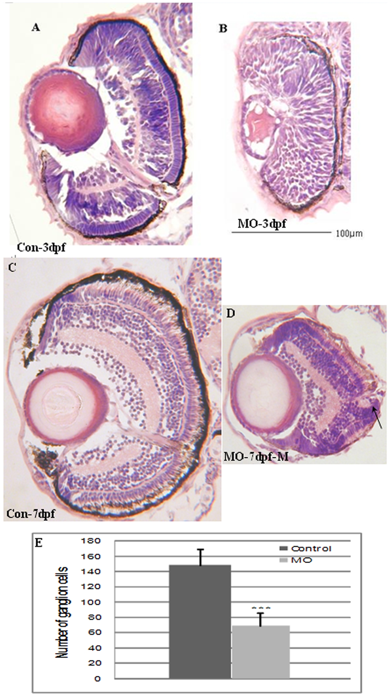
Fig. 8 Transverse sections of Atoh8-MO treated embryo retina.
(A,C) Control eyes at 3dpf and 7dpf, respectively. (B,D) Eyes of MO-treated fish at 3dpf and 7dpf, respectively. The section shows that the retina did not develop normal neuronal stratification compared with the control at 3dpf. By 7pdf, lamination had formed in the treated embryos, however, the retinal size was remarkably reduced (D), and the number of retinal ganglion neurons was significantly reduced in MO fish (E). (p<0.001). Besides, the photoreceptor layer of the morphant was less elongated. In all panels, the ventral is down.
Figure Data
Acknowledgments
This image is the copyrighted work of the attributed author or publisher, and
ZFIN has permission only to display this image to its users.
Additional permissions should be obtained from the applicable author or publisher of the image.
Full text @ PLoS One