
Image
|
Figure Caption
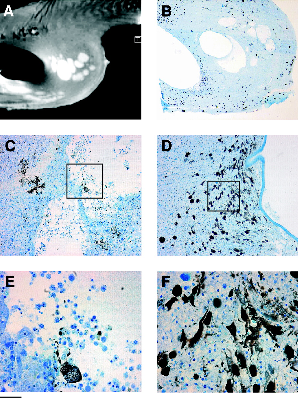
Fig. 5 Heterogeneity of the malignant abdominal tumor in transgenic zebrafish observed by (A) (MRI at 17.6 T and (B–F) after histological sectioning. Scale bars: 0.5 mm in (A, B); 0.05 mm (C, D). (E) and (F) are magnified subsampled insets of (C) and (D), respectively. Color images available online at www.liebertonline.com/zeb.
Acknowledgments
This image is the copyrighted work of the attributed author or publisher, and
ZFIN has permission only to display this image to its users.
Additional permissions should be obtained from the applicable author or publisher of the image.
Full text @ Zebrafish