
Image
|
Figure Caption
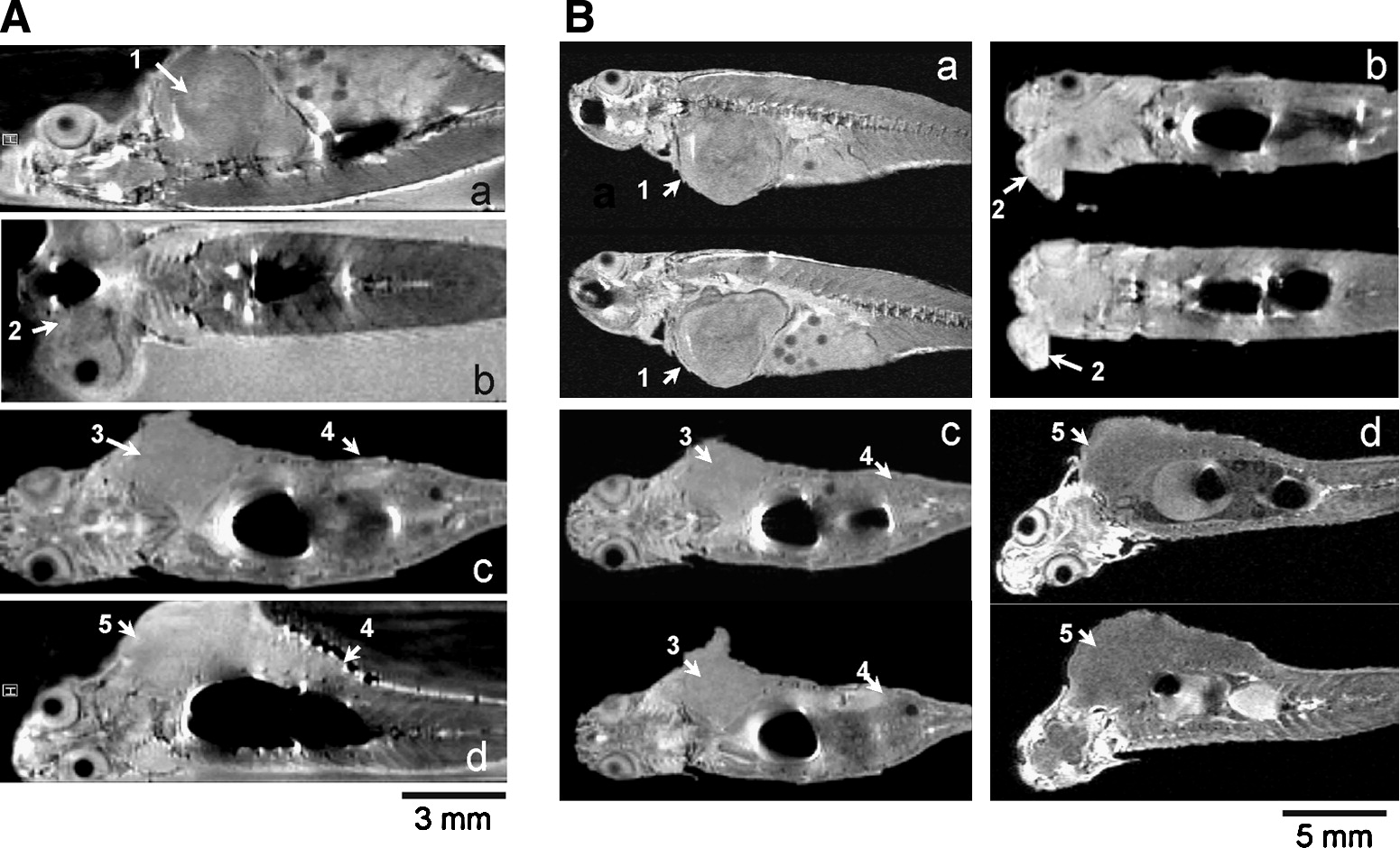
Fig. 1 Noninvasive detection of malignant tumors in transgenic mitf:Ras::mitf:GFP X p53-/- zebrafish using magnetic resonance microimaging (μMRI). Images of living (A) and freshly killed (B) adult transgenic zebrafish showing presence of tumor at various locations (a–d) in different zebrafish. Malignant tumor seen in (1) trunk muscles and abdomen that is penetrating into myoseptum; (2) near eye; (3) back muscles; (4) intestine; and (5) back muscle penetrating into liver and intestine.
Acknowledgments
This image is the copyrighted work of the attributed author or publisher, and
ZFIN has permission only to display this image to its users.
Additional permissions should be obtained from the applicable author or publisher of the image.
Full text @ Zebrafish