
Image
|
Figure Caption
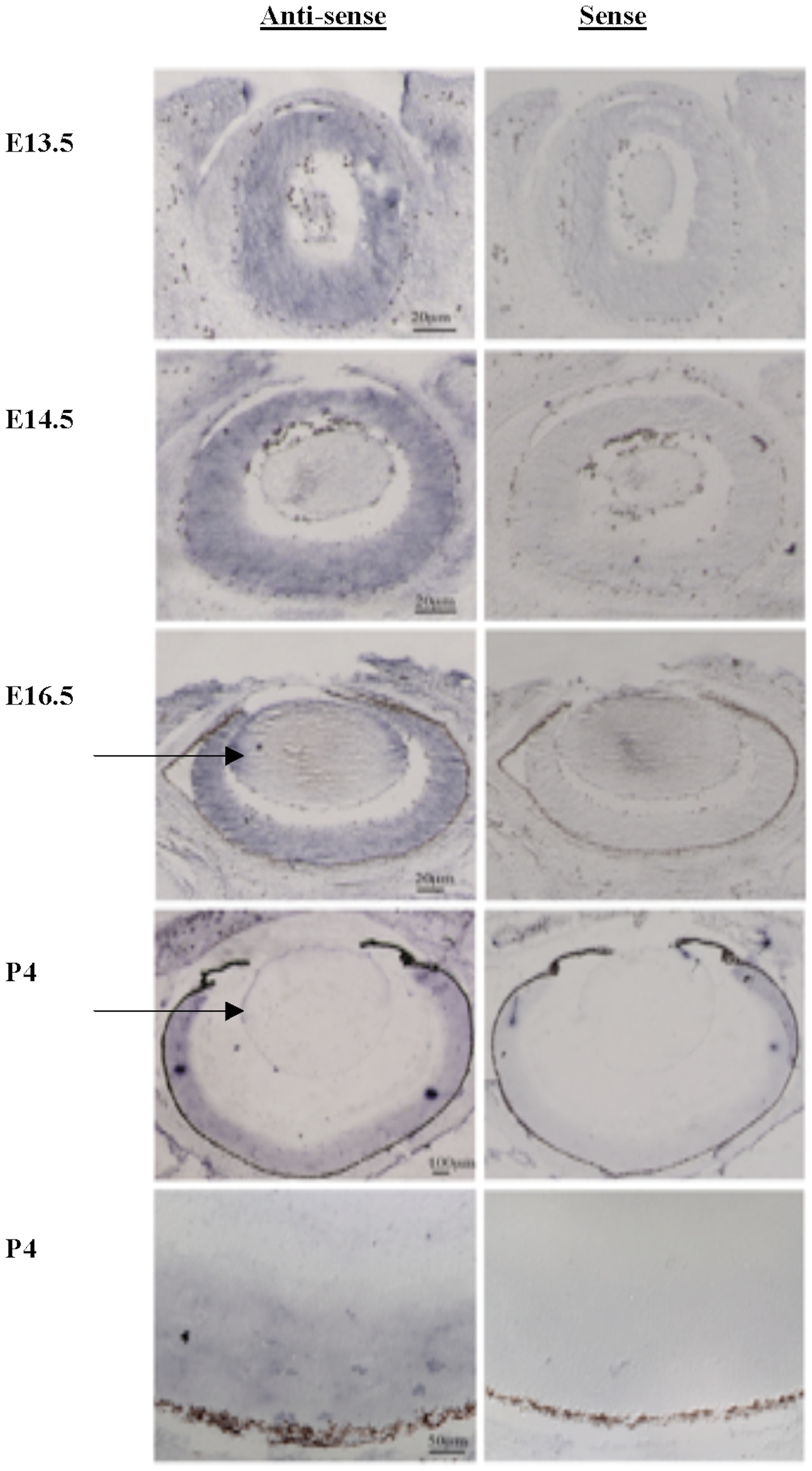
Fig. 2 In-situ hybridization shows strong expression of TMX3 in the developing murine eye.
Fig. 2. In-situ hybridization using antisense and sense riboprobes for TMX3, showing expression in the murine developing eye at E13.5, E14.5, E16.5 and P4. An arrow points to the labeling of the lens epithelium at E16.5 and P4 with the TMX3 antisense probe.
Acknowledgments
This image is the copyrighted work of the attributed author or publisher, and
ZFIN has permission only to display this image to its users.
Additional permissions should be obtained from the applicable author or publisher of the image.
Full text @ PLoS One