
|
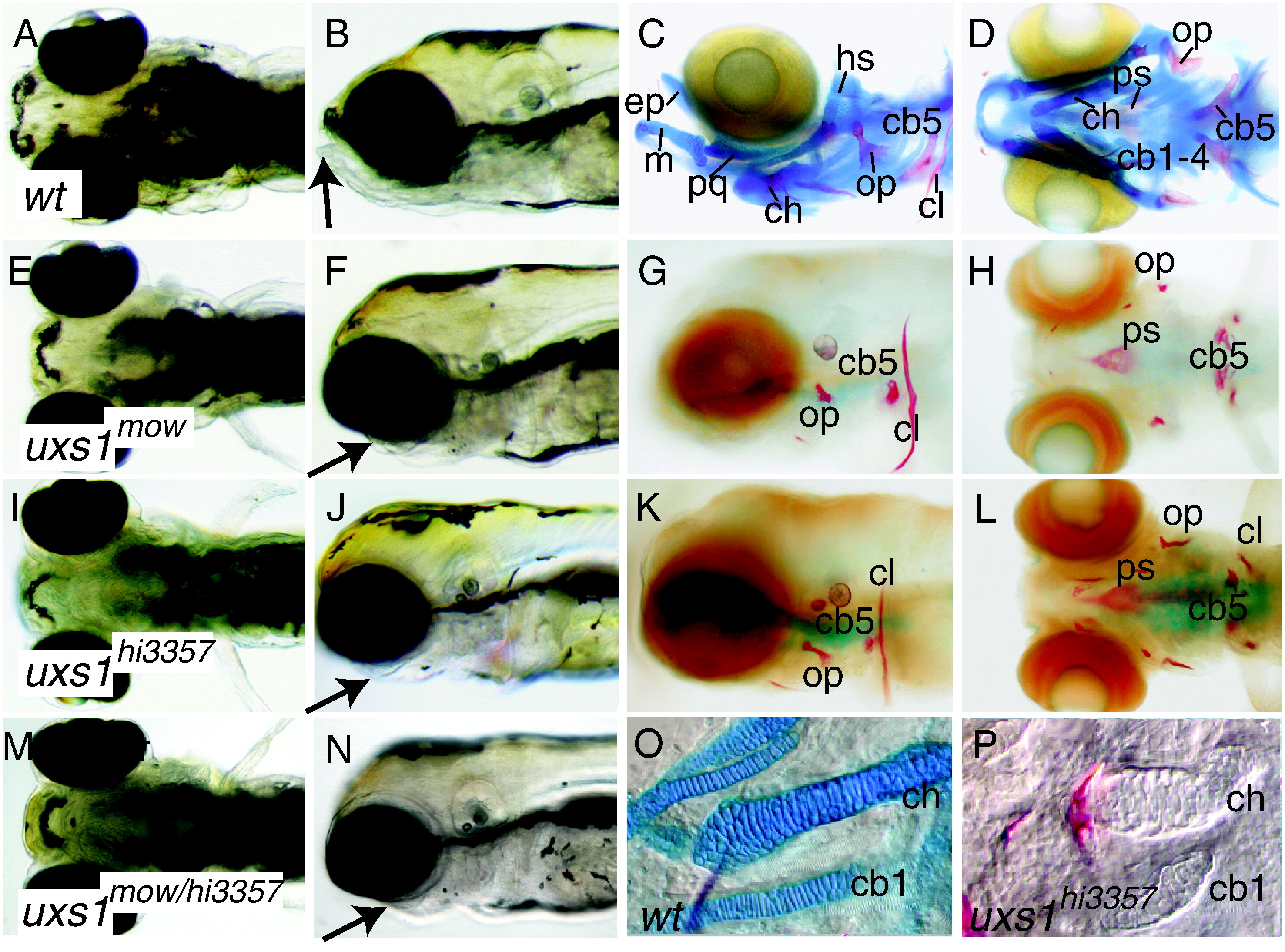
Fig. 1 Craniofacial and skeletal phenotypes of zebrafish larvae. Ventral and lateral views of live (A, B, E, F, I, J, M, N) and Alcian blue-, Alizarin red-stained (C, D, G, H, K, L, O, P) animals. Compared to wild types (A, B), mutant animals (E,F moww60 allele; I, J hi3357 allele) had reduced lower jaws (arrows) at 5 dpf. Reduced lower jaw (arrow) in moww60/hi3357 double heterozygotes (M, N) showed failure of complementation. Alcian blue and Alizarin red staining for cartilage (blue) and bone (red) revealed the lack of cartilage and reduced bones in mutants (G, H, K, L) compared to wild type (C, D) at 7 dpf. Nomarski optics on dissected pharyngeal skeletons suggested that mutant cartilages (P) condensed in the same areas as wild types (O), but did not secrete Alcian blue-positive matrix. Abbreviations: cb1-5, ceratobranchials 1 to 5; ch, ceratohyal; cl, cleithrum; ep, ethmoid plate; hs, hyosymplectic; m, Meckel′s cartilage; op, opercle; pq, palatoquadrate; ps, parasphenoid.
Reprinted from Developmental Biology, 341(2), Eames, B.F., Singer, A., Smith, G.A., Wood, Z.A., Yan, Y.L., He, X., Polizzi, S.J., Catchen, J.M., Rodriguez-Mari, A., Linbo, T., Raible, D.W., and Postlethwait, J.H., UDP xylose synthase 1 is required for morphogenesis and histogenesis of the craniofacial skeleton, 400-415, Copyright (2010) with permission from Elsevier. Full text @ Dev. Biol.