
|
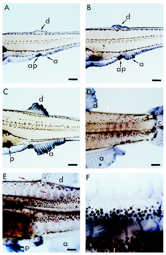
Fig. 3 Figure 3. Expression of apoE during dorsal, anal, and pelvic fins morphogenesis and during scale development. Lateral view, anterior to the left of A: 19-day, 4.8 mm B: 22-day, 5.7 mm ; C: 24-day, 6.3 mm; D: 30-day, 8.5 mm; E: 40-day, 10 mm; F: 50-day, 10.8 mm larvae. A-C: Since the embryonic stage, apoE is always expressed in the median epithelial fin fold. During the larval stages, apoE is expressed in the anal (a) and dorsal (d) fins which emerge from the fin fold, and in the pelvic fin buds (red arrow in B and (p) in C). Note that apoE is also strongly expressed in the anal papilla (ap). D: Later, apoE expression disappears in the epidermis where the median fin fold has completely regressed (white arrow), while the fins are still expressing apoE. ApoE transcripts are also detected as the scale papillae form starting in the caudal part of the trunk (red arrow). E-F: As the scale papillae develop, the domain of apoE-expressing cells becomes restricted to the posterior periphery of the scale (E : red arrow in the ventral part of the larva, and F). a, anal fin; ap, anal papilla; c, caudal fin; d, dorsal fin; p, pelvic fin. Scale bar = 200 m.