
|
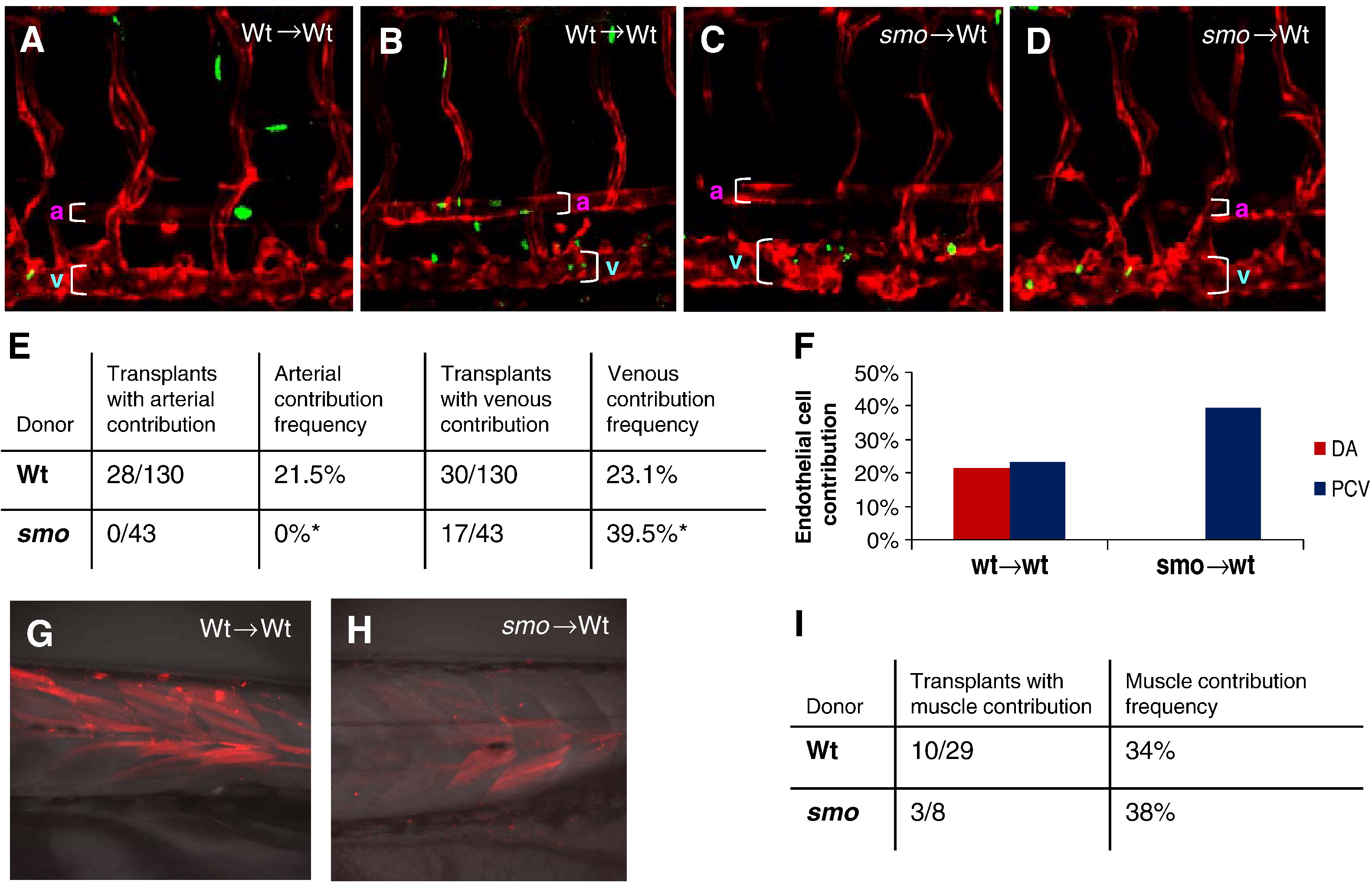
Fig. 7 Hh signaling acts cell-autonomously to induce arterial cell formation by inhibiting venous cell development. (A, B) Confocal microscopic analysis revealing the presence of EGFP-positive donor cells derived from wild-type Tg(fli:EGFP-nuc) embryos in the dorsal aorta and the posterior cardinal vein of wild-type host embryos Tg(flk:DsRed) at 72 hpf, when the dorsal aorta is completely separated from the cardinal vein. (C, D) Confocal optics showing the presence of EGFP-positive smo-null cells derived from heterozygous crosses of smo mutants [smo -/+; Tg(fli:EGFP-nuc)] in the posterior cardinal vein in wild-type host embryos Tg(flk:DsRed) at 72 hpf. (E) Data table quantitating transplantation results. A ratio of hosts with donor-derived arterial (or venous) cells to all host embryos with donor cells in any tissue was used to determine arterial (or venous) contribution frequency. (F) Bar chart depicting arterial or venous contribution frequency. (G, H) Fluorescent optics showing transplantation of fluorescein-dextran injected donor cells into wild-type hosts. Both wild-type and smo donors give rise to skeletal muscle tissue. (I) Data table quantitating transplantation results of fluorescein-dextran injected hosts. A ratio of hosts with donor-derived muscle cells to all host embryos was used to determine muscle contribution frequency.
Reprinted from Developmental Biology, 341(1), Williams, C., Kim, S.H., Ni, T.T., Mitchell, L., Ro, H., Penn, J.S., Baldwin, S.H., Solnica-Krezel, L., and Zhong, T.P., Hedgehog signaling induces arterial endothelial cell formation by repressing venous cell fate, 196-204, Copyright (2010) with permission from Elsevier. Full text @ Dev. Biol.