
|
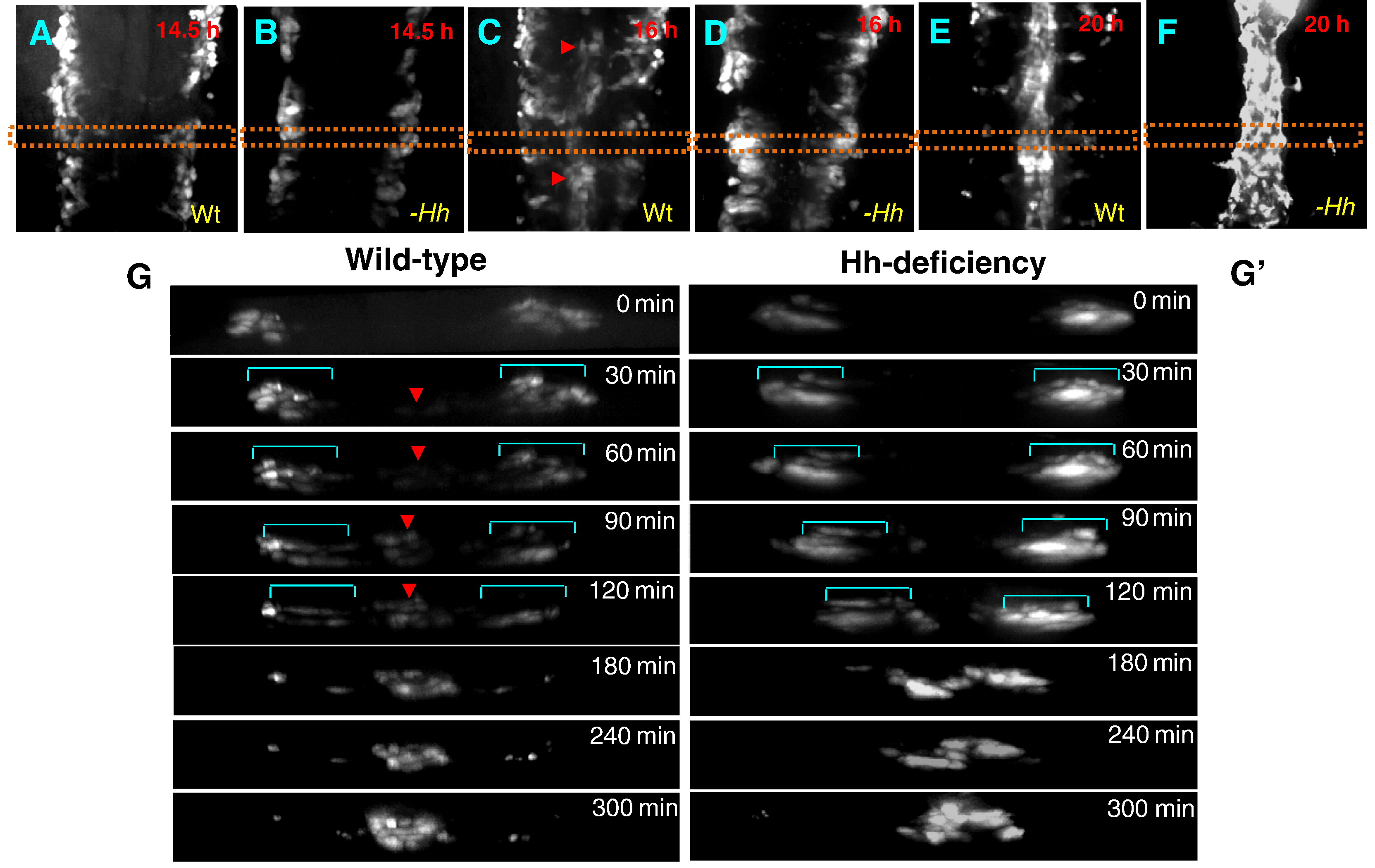
Fig. 6 The first wave of angioblast migration is diminished in embryos with Hh signaling deficiency. (A–F) Time-lapse imaging revealing sequential migration of angioblasts toward the midline from 14.5 hpf to 20 hpf in wild-type embryos (A;C;E), and cyclopamine-treated embryos (B;D;F). (G and G′) Crossover optical sections derived from the time-lapse sequence viewing angioblast migration in wild-type embryos (G), and cyclopamine-treated embryos (G′). Red arrowhead: arterial cells. Blue bracket: venous cells. Frames were captured from 300-min time-lapse sequence. Images were obtained from the dorsal side of transgenic embryos Tg(fli1:EGFP) (A–F). The 0 min time point is from the approximately 14.5 hpf stage (G;G′). Times shown in right corners represent time elapsed after the start of the sequence (G;G′). Yellow dot lines (A–F) indicate where the optical crossover sections in G and G′ were derived from.
Reprinted from Developmental Biology, 341(1), Williams, C., Kim, S.H., Ni, T.T., Mitchell, L., Ro, H., Penn, J.S., Baldwin, S.H., Solnica-Krezel, L., and Zhong, T.P., Hedgehog signaling induces arterial endothelial cell formation by repressing venous cell fate, 196-204, Copyright (2010) with permission from Elsevier. Full text @ Dev. Biol.