
|
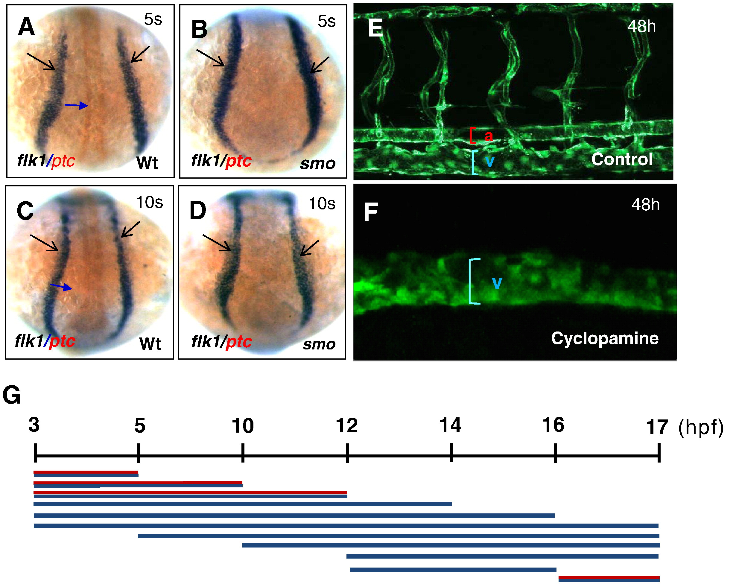
Fig. 4 Temporal development stages when Hh signaling affects artery–vein specification. (A–D) Dorsal views displaying comparable levels of flk1-positive angioblasts in wild-type embryos (A, C) and smo mutants (B, D). (E, F) Confocal microscopy analysis revealing the normal size of the dorsal aorta, the posterior cardinal vein and intersomitic vessels in control embryos (E), and an expanded posterior cardinal vein in cyclopamine-treated embryos (F). (G) Schematic representation depicting the temporal activity of cyclopamine in artery–vein specification. Embryos were incubated in 50 μM cyclopamine during a series of developmental window. Red and blue line: artery and vein. Blue line: vein. 5 s:5-somite stage. 10 s:10-somite stage.
Reprinted from Developmental Biology, 341(1), Williams, C., Kim, S.H., Ni, T.T., Mitchell, L., Ro, H., Penn, J.S., Baldwin, S.H., Solnica-Krezel, L., and Zhong, T.P., Hedgehog signaling induces arterial endothelial cell formation by repressing venous cell fate, 196-204, Copyright (2010) with permission from Elsevier. Full text @ Dev. Biol.