
|
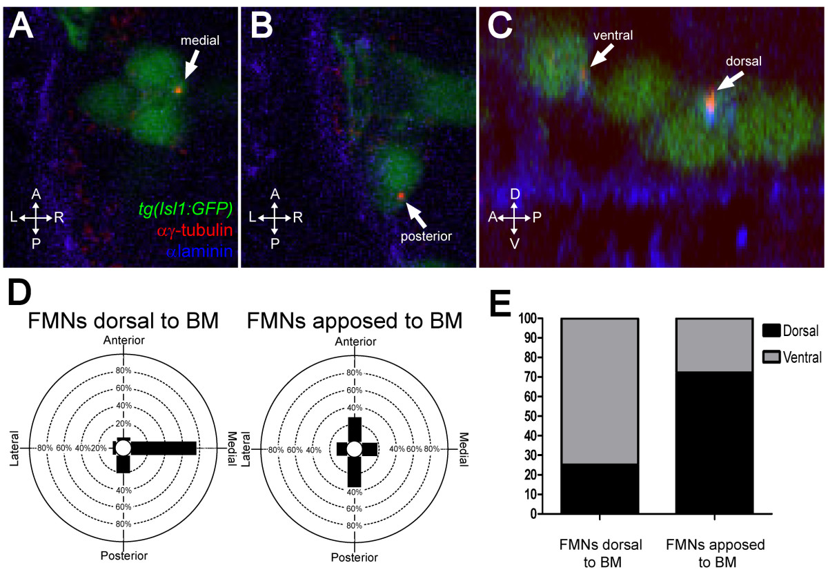
Fig. 2 FBMNs undergo a change in centrosome orientation upon contact with the Laminin-containing ventral basement membrane. (A, B) Single XY sections of 24-hpf embryos stained for Laminin (blue) and γ-tubulin (red) reveal that FBMNs that have not yet contacted the basement membrane (BM) (A) mostly orient their centrosomes medially (arrow) while FBMNs that are apposed to the BM (B) orient their centrosomes anteriorly or posteriorly (arrow). (D) Percentage of FBMNs dorsal to the BM whose centrosomes are found in the specified quadrant versus FBMNs apposed to the BM. (C) Reconstructed YZ sections of these same embryos reveal that centrosomes in FBMNs that are dorsal to the basement membrane are mostly in the ventral half of the cell while in FBMNs that are apposed to the basement membrane the centrosomes are mostly dorsal (arrows). (E) Percentage of FBMNs dorsal to the BM whose centrosomes are found in the dorsal or ventral halves versus FBMNs apposed to the BM.