
Image
|
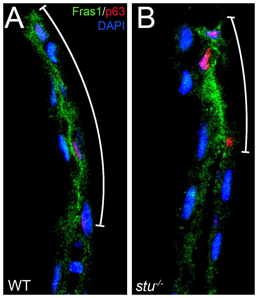
Figure Caption
Fig. S5 Fras1 distribution is compromised in sturgeon mutant fins. Transverse sections of WT (A) or stu-/- (B) posterior medial fins at 32 hpf, fluorescently immunostained for Fras1 (green), p63 (pink) and DAPI (blue). The extent of relative proximal extension of Fras1 protein appears reduced in stu-/- embryos (B) compared to WT (A). Extent of Fras1 staining is delineated by adjacent white line. Note that in the mutant, levels of Fras1 protein in the smaller domain appear correspondingly higher.
Acknowledgments
This image is the copyrighted work of the attributed author or publisher, and
ZFIN has permission only to display this image to its users.
Additional permissions should be obtained from the applicable author or publisher of the image.
Full text @ PLoS Genet.