
Image
|
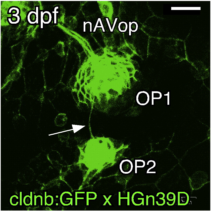
Figure Caption
Fig. S2 Sensory axons follow the budding structure. Single confocal image of a 3-dpf embryo doubly transgenic for cldnb:gfp and HGn39D. The OP1 and OP2 proneuromasts are connected by a sensory nerve (arrow). Scale bar: 20 μm.
Acknowledgments
This image is the copyrighted work of the attributed author or publisher, and
ZFIN has permission only to display this image to its users.
Additional permissions should be obtained from the applicable author or publisher of the image.
Reprinted from Developmental Biology, 340(2), Wada, H., Ghysen, A., Satou, C., Higashijima, S.I., Kawakami, K., Hamaguchi, S., and Sakaizumi, M., Dermal morphogenesis controls lateral line patterning during postembryonic development of teleost fish, 583-594, Copyright (2010) with permission from Elsevier. Full text @ Dev. Biol.