
|
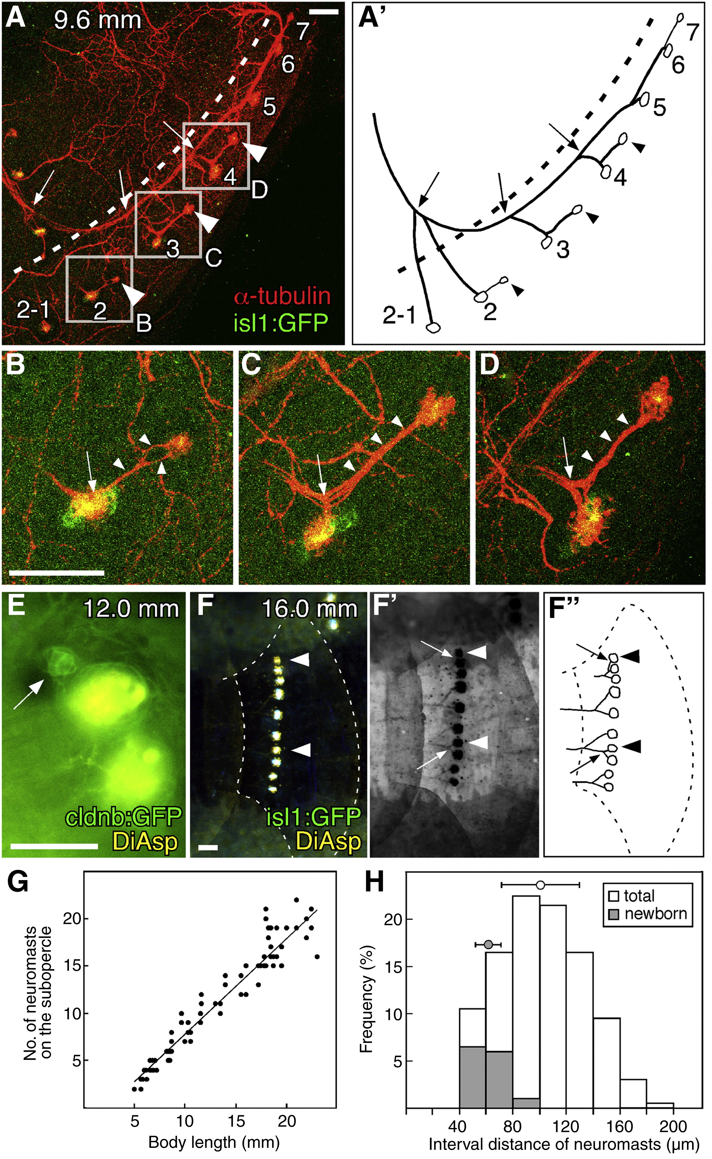
Fig. 8 Development of the pedigree-like architecture of the opercular lateral line. (A) isl1:gfp juvenile fish stained with anti-GFP and anti-acetylated α-tubulin antibodies. (A′) Schematic drawing of the lateral line architecture. Arrowheads indicate newborn neuromasts; arrows indicate the bifurcation points left behind by moving neuromasts; dotted lines indicate the boundary between opercle and subopercle. (B–D) Higher magnifications of the boxed regions in A. Arrowheads indicate the nerve extending from the mother neuromasts; arrows indicate bifurcations of the nerve. (E) Stitch formation on a body scale. Arrow indicates the budding structure. (F) isl1:gfp juvenile fish stained with DiAsp. Arrowheads indicate newborn neuromasts. (F′) The same image was converted to negative and monochrome to show the axonal trajectories retrogradely labeled with DiAsp. (F″) Schematic drawing of the innervation pattern in F′. Arrows indicate the nerve innervating the newborn neuromasts. (G) The number of DiAsp-positive neuromasts on the subopercle increases during postembryonic development. (H) Quantification of interval length between adjacent DiAsp-positive neuromasts. Intervals between newly born neuromasts and their mother neuromasts are indicated by gray bars. Mean ± SEM are indicated respectively by the circles and bars on top of the graph. Scale bars: 50 μm.
Reprinted from Developmental Biology, 340(2), Wada, H., Ghysen, A., Satou, C., Higashijima, S.I., Kawakami, K., Hamaguchi, S., and Sakaizumi, M., Dermal morphogenesis controls lateral line patterning during postembryonic development of teleost fish, 583-594, Copyright (2010) with permission from Elsevier. Full text @ Dev. Biol.