
|
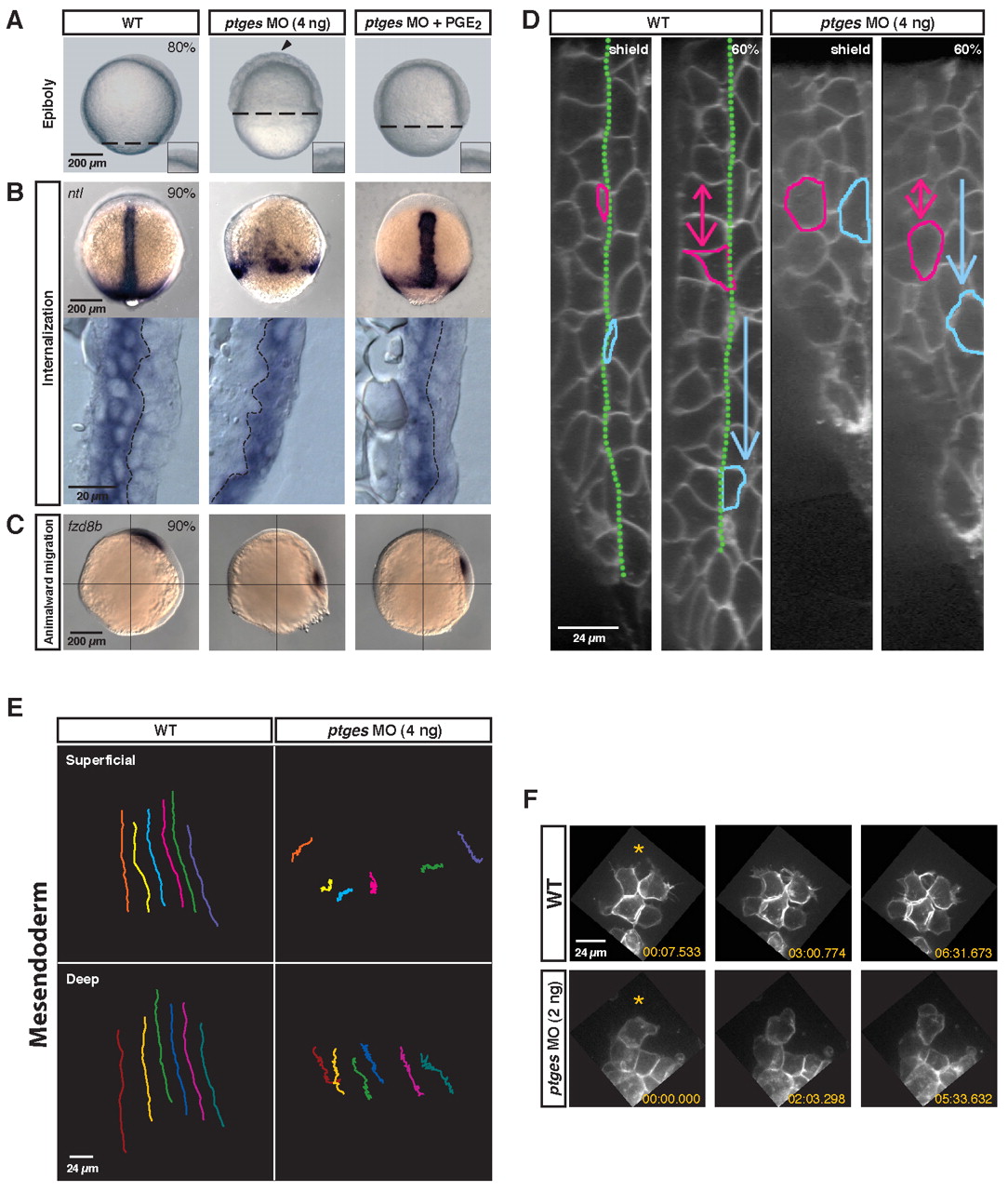
Fig. 1 Depletion of PGE2 causes global gastrulation defects. ptges MO-injected (4 ng) zebrafish embryos were evaluated for gastrulation movement defects. (A) Live embryos (80% epiboly). WT, uninjected wild type; ptges MO (4 ng), Ptges-deficient; ptges MO (4 ng) + PGE2, Ptges-deficient embryos treated with 10 mM PGE2. Dashed line indicates the progress of epiboly. Arrowhead indicates the uneven surface of the blastoderm. The inset shows the blastoderm surface at high magnification. (B) (Top) Deficit of internalized no tail-expressing axial mesodermal cells in ptges morphants (90% epiboly; dorsal views). (Bottom) Lateral views of cryosections through the dorsal side of the embryo. Yolk cell, left; external surface, right; dashed line, germ layer boundary. (C) Migration of fzd8b-expressing prechordal mesoderm cells towards the animal pole (90% epiboly; lateral views). (D) Confocal images (lateral view) of the dorsal midline in membrane EGFP-labeled embryos at shield stage. Yolk cell, left; external surface, right. The green line marks the boundary between the superficial (blue cell) and deep layers (pink cell) of the gastrula. Arrows indicate the vector of movement. (E) The paths of six cells in the superficial and deep layers of the mesendoderm in wild-type and ptges morphant embryos. Vegetal pole, top; animal pole, bottom. (F) Confocal images of prechordal mesoderm cells (70% epiboly). Three frames from a time-lapse movie are shown. Orange asterisks are towards the animal pole.