
Fig. 3
- ID
- ZDB-IMAGE-100422-55
- Publication
- Schonthaler et al., 2010 - The zebrafish mutant bumper shows a hyperproliferation of lens epithelial cells and fibre cell degeneration leading to functional blindness
- All Figures
- Figures for Schonthaler et al., 2010
|
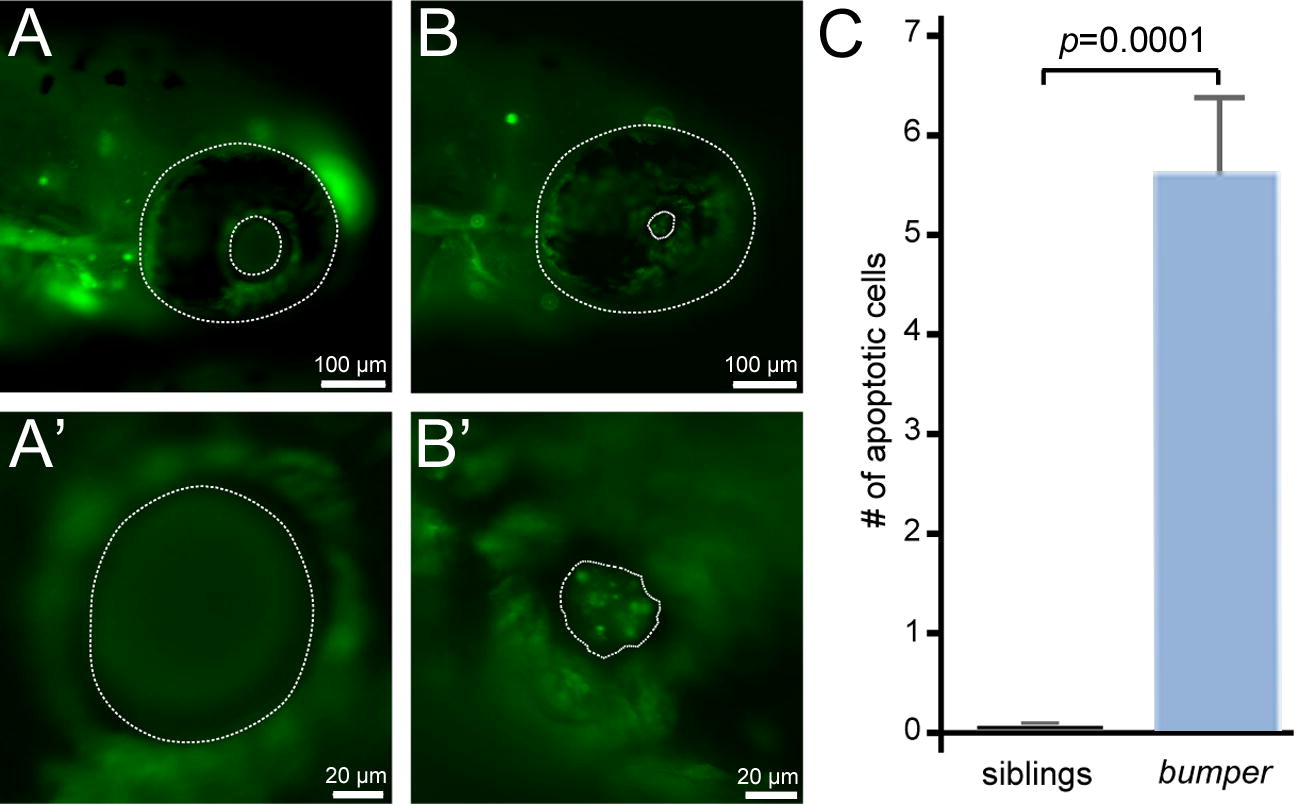
Fig. 3 Mutant bum-/- lenses display significantly increased levels of apoptotic cell death in the anterior lens region at 4 dpf. (A, B) External views of the heads (A, B) and eyes (A′, B′) of 4 dpf sibling (wild-type, wt; A, A′) and bum-/- larvae (B, B′) showing Acridine Orange fluorescence to reveal cells undergoing apoptosis . The outer margins of the eyes (outer dotted lines in A and B) and the inner margins of the pupils (inner dotted lines in A and B; dotted lines in A′ and B′) are indicated. Note the reduced and irregular pupil diameter in the bum-/- mutant larvae. While the wild-type lenses do not show Acridine Orange-positive cells (A′), mutant lenses regularly contain numerous fluorescent cells (B). (C) Statistical analysis of the number of apoptotic cells in 4 dpf sibling (left) and bum-/- mutant larvae (right) showing highly significant numbers of apoptotic cells in the mutant lenses (but not in the wild-type), which suggests a mechanism for the regression of the lens tumours in bum-/-. Error bars represent the standard error of the mean (SEM).
Reprinted from Mechanisms of Development, 127(3-4), Schonthaler, H.B., Franz-Odendaal, T.A., Hodel, C., Gehring, I., Geisler, R., Schwarz, H., Neuhauss, S.C., and Dahm, R., The zebrafish mutant bumper shows a hyperproliferation of lens epithelial cells and fibre cell degeneration leading to functional blindness, 203-219, Copyright (2010) with permission from Elsevier. Full text @ Mech. Dev.