
|
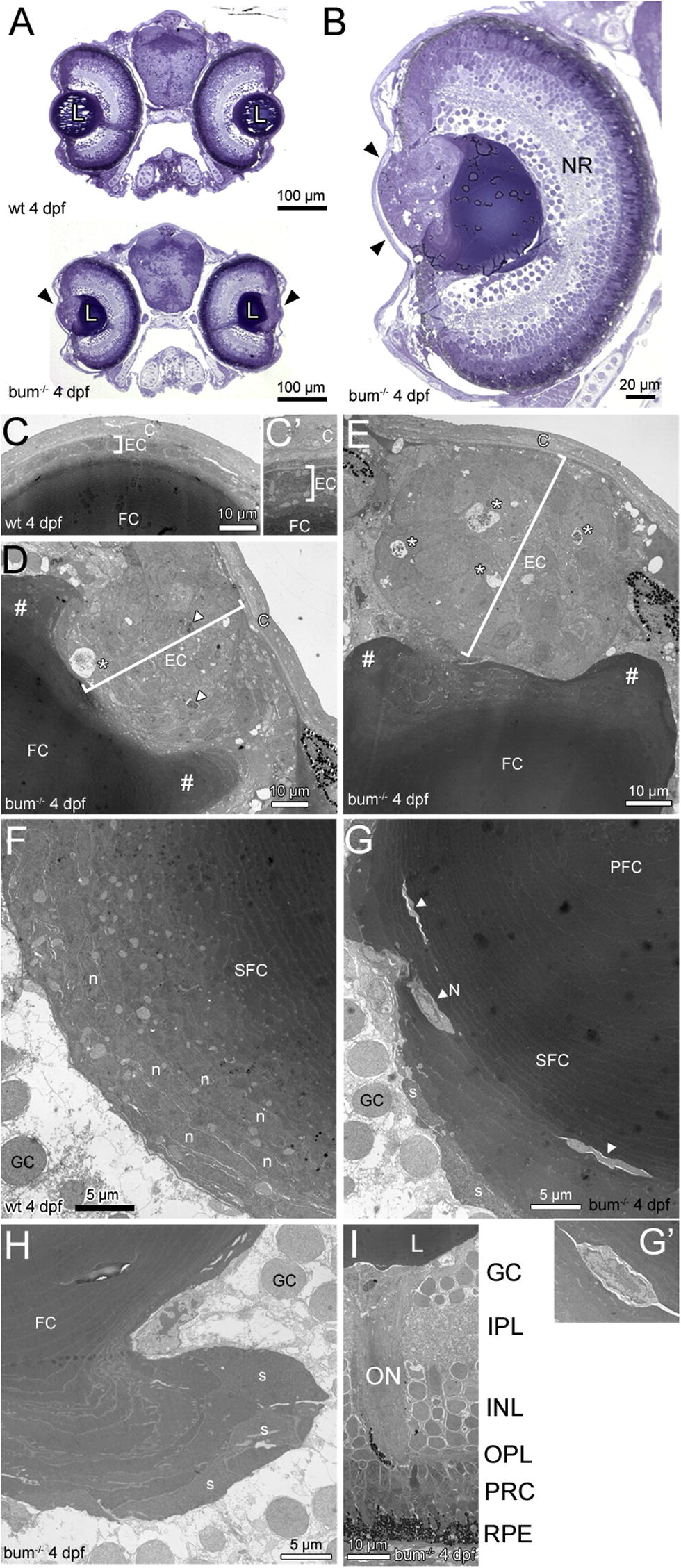
Fig. 2 The lens in 4 dpf bum-/- zebrafish shows a massive hyperproliferation of the anterior epithelium and the onset of a maldifferentiation of secondary fibre cells. (A, B) Toluidine blue-stained transversal semi-thin sections through the central part of the eyes of 4 dpf sibling (wild-type, wt; top picture in A) and bum-/- zebrafish larvae (bottom picture in A and B). (A) Cross-sections of entire heads, (B) A higher magnification of an individual eye. Note the hyperproliferation of the anterior lens epithelium in the mutant (black arrowheads), which does not affect the differentiation of the primary and early developing secondary fibre cells (collectively termed “FC” here as they cannot be differentiated at the magnification shown in B). The overall morphology of the head (A) and eye (B), including the development of the layers of the neural retina (NR), are normal in bum-/- mutant larvae. (C–I) Thin-section electron microscopy images of transversal sections through the central part of the eyes of 4 dpf bum-/- zebrafish larvae. (C–E) Anterior part of the eyes of 4 dpf sibling (C; higher magnification C′) and bum-/- mutant (D, E) zebrafish larvae. Note that while wild-type larvae consistently have a monolayered lens epithelium (brackets in C, C′), the mutants display a massively hyperproliferating lens epithelium (brackets in D, E). Moreover, the mutant lens epithelium frequently comprises pyknotic nuclei (arrowheads in D) and empty spaces containing cell debris (asterisks in D and E). The # signs indicate the massive deformation of the fibre mass near the anterior lens pole. (F, G) Bow regions posterior of lens equator encompassing the early differentiating lens secondary fibre cells in 4 dpf sibling (F) and bum-/- mutant (G) zebrafish larvae. In the mutant lenses note the presence of cytoplasm with a much lighter appearance (arrowheads and inset G′), sometimes still containing nuclei (N, inset), embedded in layers of secondary fibre cells that appear to have differentiated normally. Also note the swelling of some of the outermost fibre cells (s), which is however not yet as pronounced as at later developmental stages (see Fig. 4). (H) Lobe-like protrusion of secondary lens fibre cells near the posterior pole of a bum-/- mutant lens. Note that some of the fibre cells, particularly in the outermost layers, appear to be swollen (s). (I) Part of the central retina showing normal development of the layers of the neural retina, the optic nerve and the retinal pigment epithelium. Abbreviations: C, cornea; EC, lens epithelial cells; FC, (primary and secondary) fibre cells; GC, retinal ganglion cells; INL, inner nuclear layer; IPL, inner plexiform layer; L, lens; N, remaining nucleus in a late-stage differentiating secondary lens fibre cell; n, nuclei in a early-stage differentiating secondary lens fibre cells; NR, neural retina; ON, optic nerve; OPL, outer plexiform layer; PFC, primary lens fibre cells; PRC, photoreceptor cells; RPE, retinal pigment epithelium; SFC, secondary lens fibre cells.
Reprinted from Mechanisms of Development, 127(3-4), Schonthaler, H.B., Franz-Odendaal, T.A., Hodel, C., Gehring, I., Geisler, R., Schwarz, H., Neuhauss, S.C., and Dahm, R., The zebrafish mutant bumper shows a hyperproliferation of lens epithelial cells and fibre cell degeneration leading to functional blindness, 203-219, Copyright (2010) with permission from Elsevier. Full text @ Mech. Dev.