
|
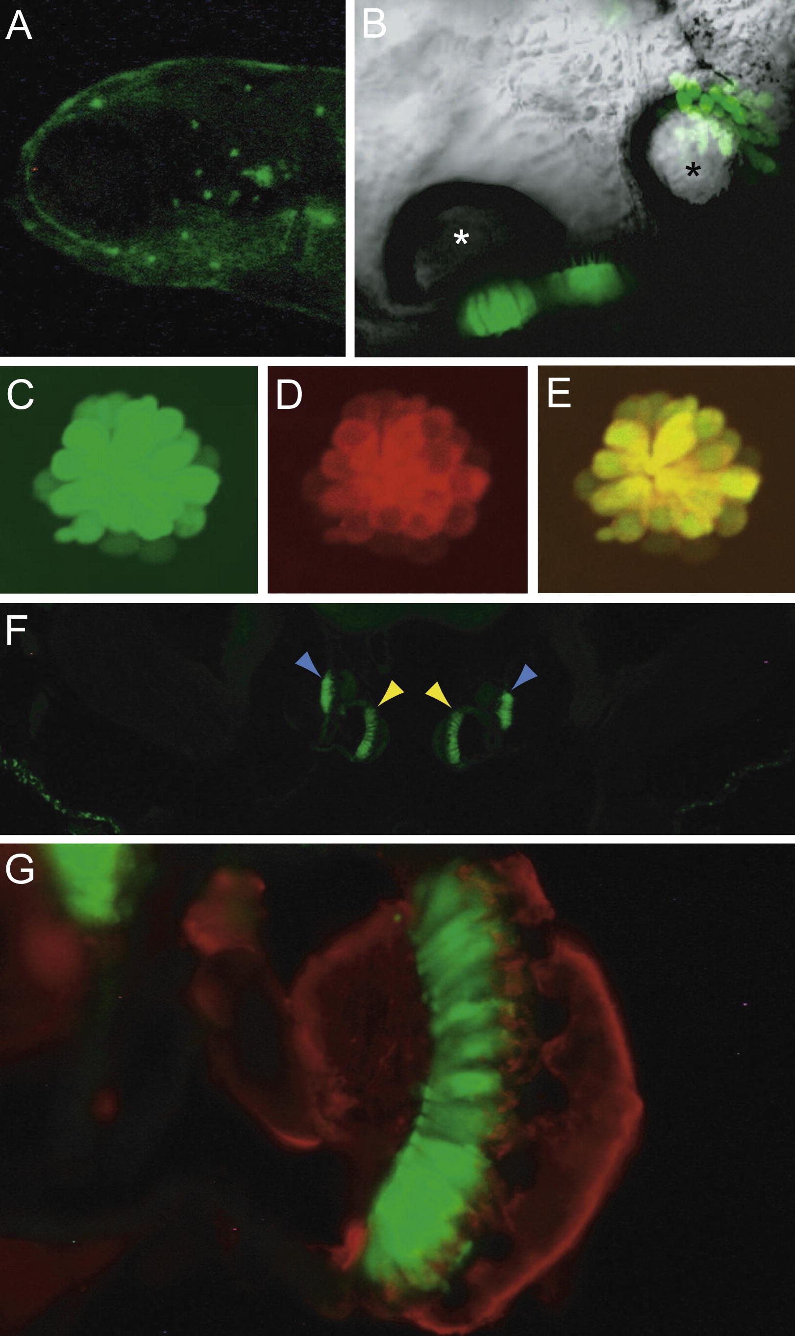
Fig. 2 Cellular distribution of GFP in the Ppv3a-3 transgenic line. (A) A fluorescent image of a 5-dpf larval head demonstrates labeling of neuromasts in the anterior lateral line. (B) In a ventral view of an otocyst in a 5-dpf larva, sensory epithelia containing GFP-positive hair cells are located beneath the two otoliths. The anterior otolith is marked with a white asterisk, the posterior otolith with a black one. (C) Transgenically expressed GFP marks hair cells in a 5-dpf larval neuromast. (D) Labeling with FM4-64, a fluorophore that passes through functional mechanotransduction channels, reveals the complement of mature hair cells. (E) The merged image of GFP and FM4-64 signals confirms that the transgenic label marks all the functional hair cells. (F) A transverse section of the head of an adult fish portrays the GFP-positive sensory maculae of the medial sacculi (yellow arrowheads) and the lateral lagenae (blue arrowheads). (G) A higher-magnification image of one sacculus shows GFP-positive hair cells (green) and nerve fibers labeled with an antiserum against neurofilaments (red).
Reprinted from Gene expression patterns : GEP, 10(2-3), McDermott, B.M., Asai, Y., Baucom, J.M., Jani, S.D., Castellanos, Y., Gomez, G., McClintock, J.M., and Hudspeth, A.J., Transgenic Labeling of Hair Cells in the Zebrafish Acousticolateralis System, 113-118, Copyright (2010) with permission from Elsevier. Full text @ Gene Expr. Patterns