
|
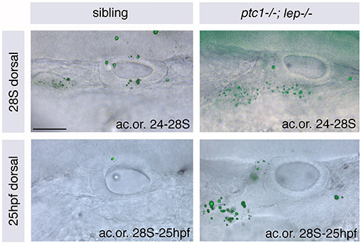
Fig. S2 There is no increase in cell death in DL regions of ptc1-/-; lep-/- otic vesicles compared with those of siblings. Otic vesicles of wild-type and ptc1-/-; lep-/- embryos stained with Acridine Orange (ac.or.) between 24 and 28S (top) and 28S and 25 hpf (bottom) to reveal regions of cell death. Fluorescent images showing Acridine Orange stain are overlaid onto DIC images of live ears. There is no increase in cell death in the ptc1-/-; lep-/- otic vesicle itself. However, there is a significant increase anteroventral and lateral to the otic vesicle, which is likely to include the forming statoacoustic ganglion. Dorsal views; anterior to left, medial to top. Scale bar: 50 μm.