
|
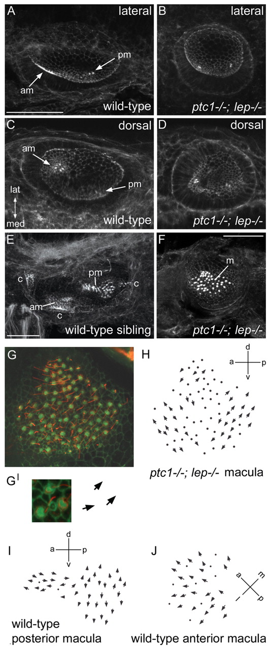
Fig. 4 Otic vesicles of ptc1-/-; lep-/- embryos contain a single medial macula with double posterior polarity, which originates as two separate domains of hair cells. (A-F) Merged confocal z-stacks of otic vesicles at 33 hpf (A-D) and 3 dpf (E,F) stained with FITC-phalloidin. Hair cell bundles appear as bright dots. Two separate sensory patches are seen in ptc1-/-; lep-/- ears (B,D), as in wild-type ears, but both arise in a medial position like the posterior patch in wild-type ears (compare C with D). By 3 dpf, the patches have merged to form a single macula in ptc1-/-; lep-/- (F). (G) Merged confocal z-stack of a 70 hpf ptc1-/-; lep-/- macula stained with FITC-phalloidin (cuticular plate/stereocilia; green) and anti-acetylated tubulin antibody (kinocilia; red). (G′) Enlarged image of three hair cells, showing the planar polarity of each hair cell (arrows). (H) Polarity map of the ptc1-/-; lep-/- medial macula shown in G. Additional examples are shown in Fig. S3 in the supplementary material. Hair bundles point away from a midline in both the anterior and posterior halves of the macula, resembling the pattern seen in wild-type posterior maculae. (I,J) Polarity maps of wild-type posterior and anterior maculae for comparison. (A,B,E,F) Lateral views; anterior to left, dorsal to top. (C,D) Dorsal views; anterior to left, lateral to top. am, anterior macula; c, crista; lat, lateral; m, medial macula; med, medial; pm, posterior macula. Scale bars: 50 μm (bar in A applies to A-D).