
|
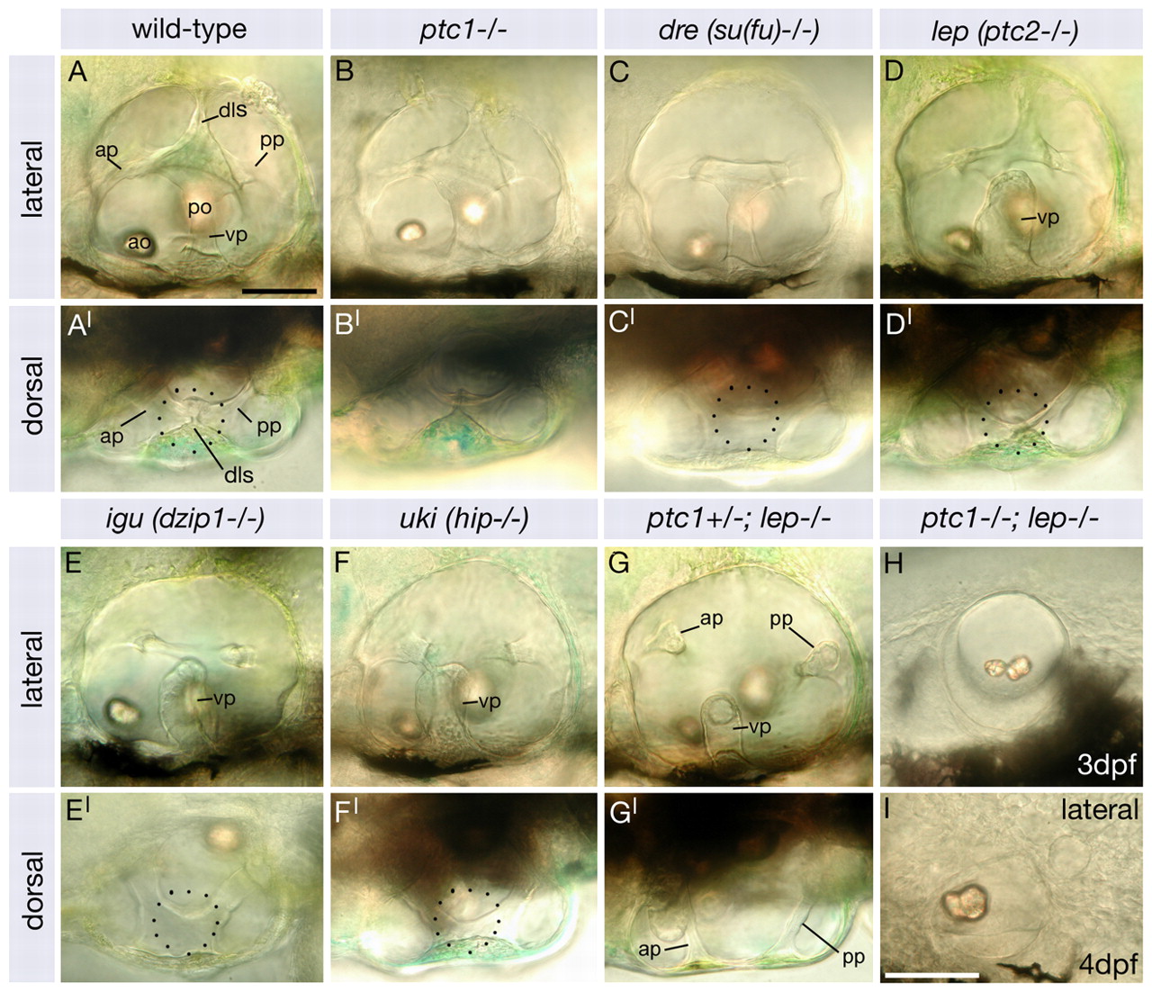
Fig. 1 Dorsolateral otic structures are lost with increasing severity of otic phenotype in a panel of Hh pathway inhibitor mutants. Images of live zebrafish inner ears taken using DIC optics at 4 dpf, except in H (3 dpf). (A-I) Lateral views; anterior to left, dorsal to top. (A′-G′) Dorsal views; anterior to left, lateral to bottom. (A,A′) Wild-type inner ear showing the three pillars around which the semicircular canals form, the dorsolateral septum and the two otoliths. (B,B′) ptc1-/- ears are normal. (C,C′) The dls is absent in dre. (D,D′) In lep the dls and ventral canal pillar (vp) are abnormal. (E-F′) In igu and uki the dls is absent and the ventral canal projection is abnormal. (G,G′) In ptc1-/-; lep+/- all three canal projections are reduced and the dls is absent. (H,I) In ptc1-/-; lep-/- embryos the ear is small, the otoliths fuse and no canal projections or dls form. Dotted circle denotes dls (A′,D′) or region where it should form (C′,E′,F′). Scale bars: 50 μm (bar in A applies to A-H). ap, anterior semicircular canal pillar; ao, anterior otolith; dls, dorsolateral septum; po, posterior otolith; pp, posterior semicircular canal pillar; vp, ventral semicircular canal pillar.