
|
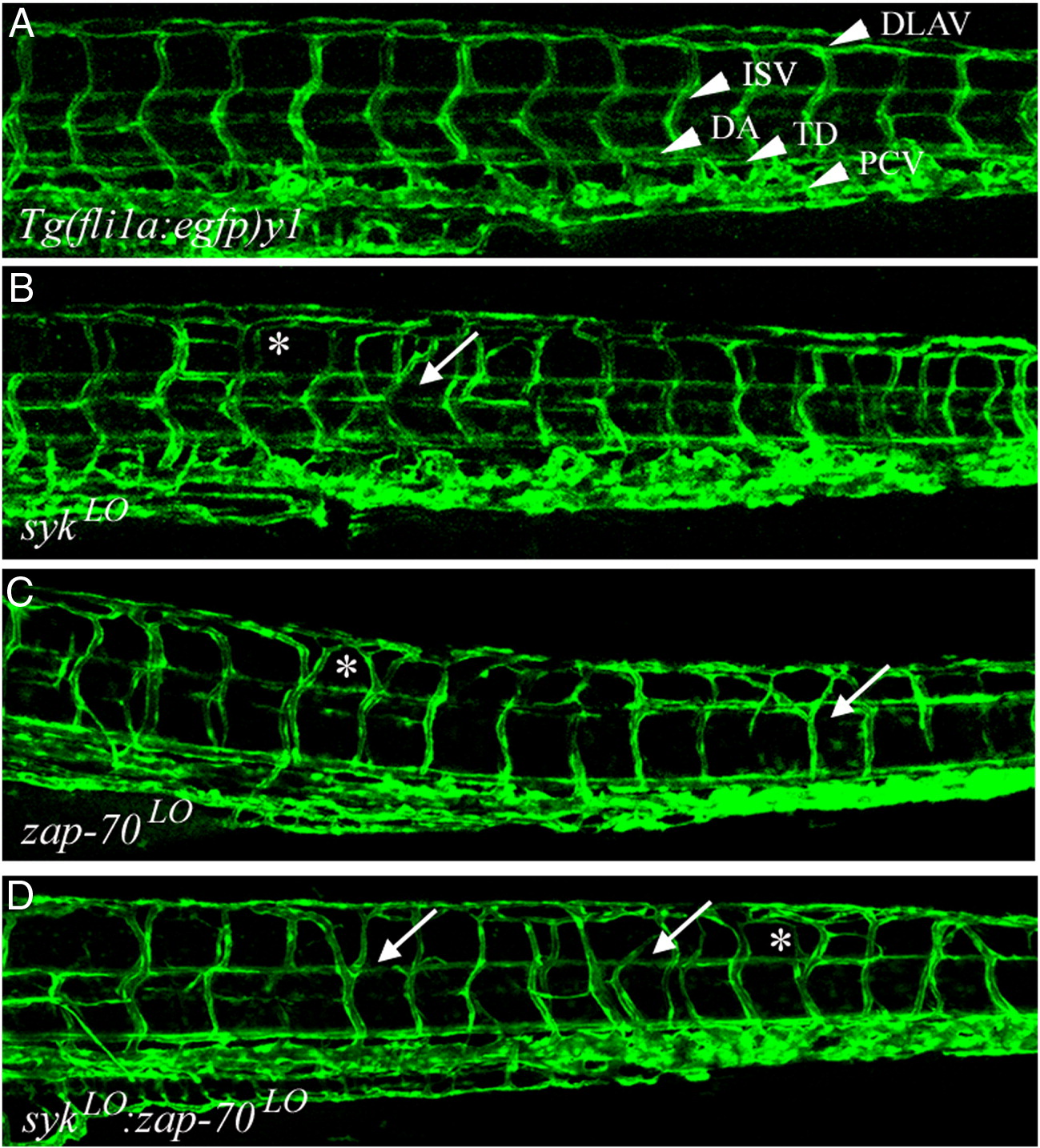
Fig. 2 sykLO and zap-70LO morphants display late stage ISV patterning defects. (A) The trunk of 5 dpf wild-type Tg(fli1a:EGFP)y1 embryos display regularly patterned ISVs between somites, with the dorsal-most ISV endothelial cell forming a clear T shape and contributing to the DLAV. In contrast, 5 dpf sykLO (B), zap-70LO (C), and sykLO:zap-70LO (D) morphants display abnormal ISV vasculature with mild patterning defects. The ventral stalk ISV endothelial cell is relatively normal, and the dorsal T-cell still contributes to the DLAV, but the branching of the cell into the T shape is abnormal (asterisks) and can occur as ventrally as midsomite (arrows). These dorsal patterning defects are more pronounced in the sykLO:zap-70LO morphants.
Reprinted from Developmental Biology, 340(1), Christie, T.L., Carter, A., Rollins, E.L., and Childs, S.J., Syk and Zap-70 function redundantly to promote angioblast migration, 22-29, Copyright (2010) with permission from Elsevier. Full text @ Dev. Biol.