
|
Fig. S1 Mutations in the lta4h Locus Result in an Extracellular Cording Phenotype, Related to Figure 2
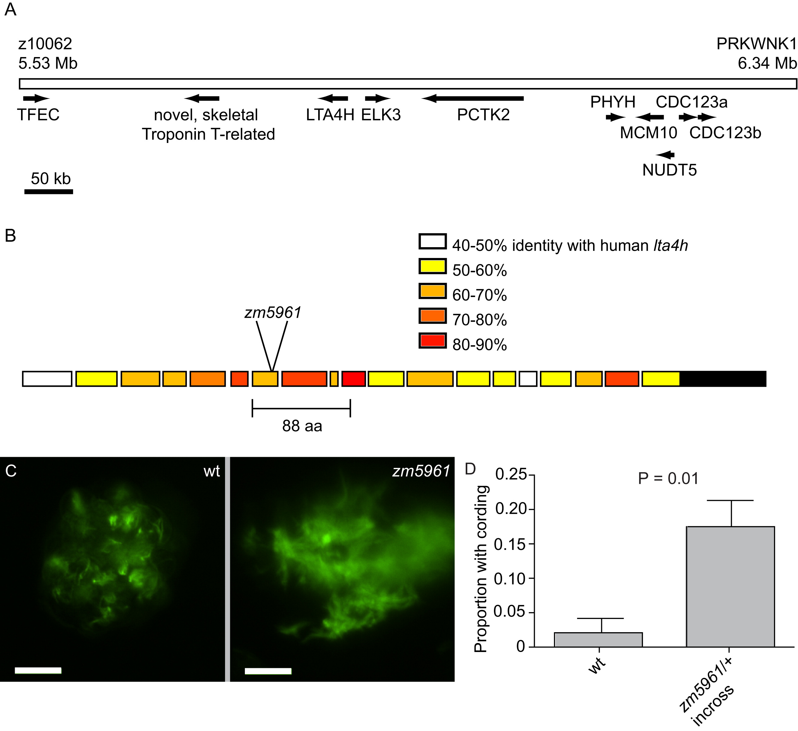
(A) Critical interval on zebrafish Chromosome 4 for fh112, bounded by closest polymorphic genetic markers. Predicted ORFs in the interval are indicated with arrows. Numbering and gene predictions based on Ensembl Zv6 assembly (www.ensembl.org).
(B) Gene structure of zebrafish lta4h locus with percent identity to human lta4h color-coded. Location of the retroviral insertion in the zm5961mutant and position of its in-frame 88 amino acid deletion is indicated. (C) Fluorescence image of non-cording bacteria within a granuloma in matched sibling control animals (left) and cording bacteria in zm5961 (right) at 5 dpi with infection dose of 161 ± 33 (SD) bacteria. Scale bars, 20 μm.
(D) Mean proportion of animals with cording in three independent groups of 15-25 animals five dpi with 161 ± 33 (SD) bacteria. p = 0.01 (Student′s unpaired t test). Error bars, SEM.
Reprinted from Cell, 140(5), Tobin, D.M., Vary, J.C. Jr, Ray, J.P., Walsh, G.S., Dunstan, S.J., Bang, N.D., Hagge, D.A., Khadge, S., King, M.C., Hawn, T.R., Moens, C.B., and Ramakrishnan, L., The lta4h Locus Modulates Susceptibility to Mycobacterial Infection in Zebrafish and Humans, 717-730, Copyright (2010) with permission from Elsevier. Full text @ Cell