
|
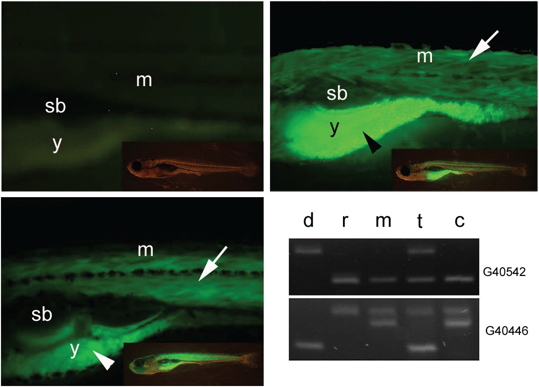
Fig. 2 Transplanted follicles develop to fertilizable eggs and produce viable offspring. (A) Non-transgenic offspring from recipient female mated with wild type male. (B) Transgenic offspring of a sibling of the donor female mated with a wild type male. (C) Transgenic offspring developing from a recipient female, which was injected with transgenic donor follicles and mated with wild type male 3 weeks after transplantation. All larvae are shown at 10 days after fertilization. Transgenic and non transgenic offspring of recipient female were analysed at 10 days post fertilization. Side view onto the trunk above the yolk ball and part of tail are shown. Fluorescence signal of the β-actin:yfp reporter construct activity is detected in the skeletal muscle and the yolk ball (arrow and arrowhead respectively). Inserts show the full view of larvae respectively. (D) Microsatellite analysis of genetic composition of donor female (d), recipient female (r), male used for crossing with recipient female (m), offspring developing from transplanted follicle (t) and sibling from recipient female (c). The identification number of microsatellite markers are indicated on the right (for details see Table 2). Abbreviations, (sb) swim bladder, (m), muscle, (y) yolk.