
|
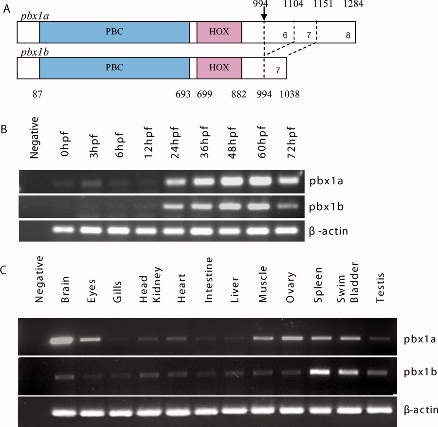
Fig. 1 Pbx1 protein structure and reverse transcriptase-polymerase chain reaction (RT-PCR) expression pattern analysis. A: Protein domains of zebrafish Pbx1a and Pbx1b. Numbers refer to amino acid residues. The location of the alternatively spliced exon is marked by an arrow. Exons 1 to 5 are common to both Pbx1 isoforms. B: RT-PCR temporal expression pattern of zebrafish pbx1a and pbx1b during early embryonic development. Sizes of the amplified products were 641 bp for pbx1a, 530 bp for pbx1b, and 200 bp for β-actin. C: Expression of pbx1a and pbx1b in adult tissues obtained from 6-month-old zebrafish.