
|
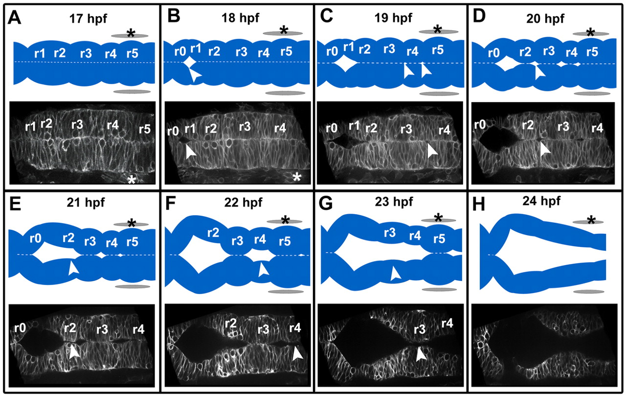
Fig. 1 The sequence of zebrafish wild-type hindbrain ventricle opening. Time course of wild-type hindbrain ventricle opening from 17 to 24 hpf. Embryos were injected with memGFP mRNA (n=12). Images (and corresponding diagrams) are a representative snapshot of the hindbrain opening at each time point. (A) At 17 hpf the neural tube is closed with visible rhombomere (r) morphology. (B) At 18 hpf the initial opening is visible at the r0/r1 boundary. (C) At 19 hpf openings surrounding r4 are visible. (D) At 20 hpf there are openings at the r2/r3 boundary and r1 has separated completely. (E) At 21 hpf opening within r2 occurs. (F) At 22 hpf separation within r4 has occurred. (G) At 23 hpf separation within r3 has occurred. (H) At 24 hpf separation within r5 occurs resulting in an open ventricle and less prominent rhombomere morphology. Asterisks indicate the ear. Arrowheads indicate regions that are separating. Anterior is to the left.