
Image
|
Figure Caption
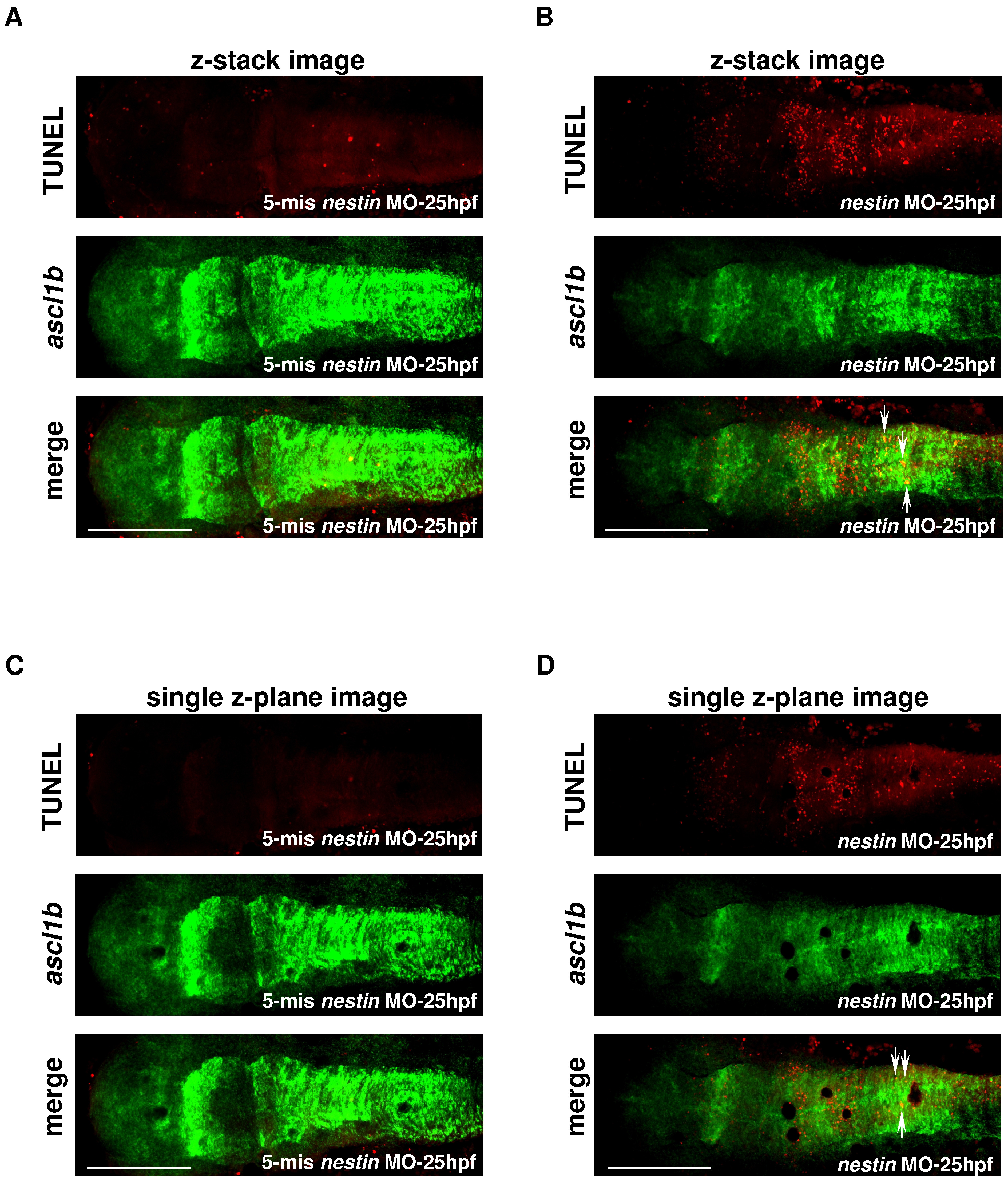
Fig. 8 Colocalization of TUNEL signals with ascl1b expression.
A and B, z-stack images and C and D, single z-plane images. Embryos were injected with (A) and (C) 5-mis nestin MO or (B) and (D) nestin MO and images were analyzed by confocal microscopy at 25 hpf. Top panels show TUNEL and middle panels, ascl1b stained by fluorescence in situ hybridization. The lower panels show merged images. Yellow dots as denoted by white arrows indicate colocalization of ascl1b with TUNEL. Bar scale: 200 μm.
Figure Data
Acknowledgments
This image is the copyrighted work of the attributed author or publisher, and
ZFIN has permission only to display this image to its users.
Additional permissions should be obtained from the applicable author or publisher of the image.
Full text @ PLoS One