
|
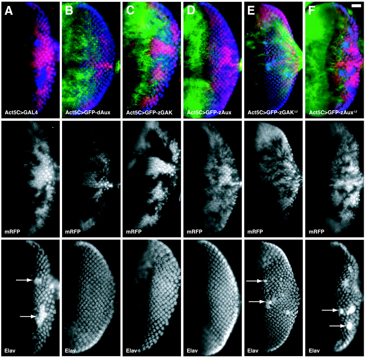
Fig. 4 Both zGAK and zAux can substitute for Drosophila auxilin. Fluorescent micrographs of Drosophila eye imaginal discs stained with αElav antibody, which labels the nuclei of the neuronal cells (blue). The expressions of zebrafish and Drosophila auxilin genes are shown in green. Homozygous dAuxF956* mutant tissues are indicated by the absence of the membrane-associated mRFP (red). Regions containing excessive Elav-positive cells are indicated by arrows. All the flies carry Act5C-GAL4, UAS-FLP on the second chromosome, and other relevant genotypes include: (A) FRT5-5Z3515, dAuxF956*/FRT5-5Z3515, GMR-src-mRFP, (B) UAS-dAux-GFP; FRT5-5Z3515, dAuxF956*/FRT5-5Z3515, GMR-src-mRFP, (C) UAS-GFP-zGAK; FRT5-5Z3515, dAuxF956*/FRT5-5Z3515, GMR-src-mRFP, (D) UAS-GFP-zAux; FRT5-5Z3515, dAuxF956*/FRT5-5Z3515, GMR-src-mRFP, (E) UAS-GFP-zGAKΔJ; FRT5-5Z3515, dAuxF956*/FRT5-5Z3515, GMR-src-mRFP, and (F) UAS-GFP-zAuxΔJ; FRT5-5Z3515, dAuxF956*/FRT5-5Z3515, GMR-src-mRFP. Scale Bar, 50 μm.