
|
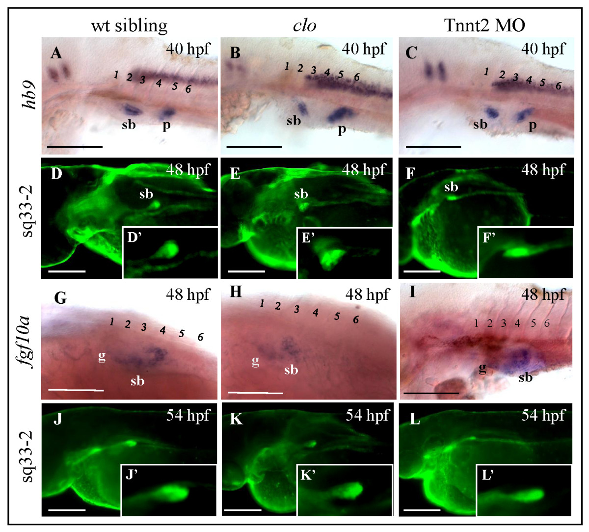
Fig. 2 Role of endothelial cells during swimbladder budding and early growth phase. (A - C) Expression of swimbladder epithelial marker hb9 in control, clo-/- mutant, and Tnnt2 morphant at the beginning of budding stage (40 hpf). Note the similar size of epithelial bud in all these embryos. (D - F) EGFP expression in control, clo-/-, and Tnnt2 morphants on Et(krt4:EGFP)sq33-2 background at 48 hpf. (D′ - F′) A 2.5x magnification of the swimbladder in (D - F). (G - I) Expression of fgf10a mesenchymal marker in swimbladders clo-/-, mutant, and Tnnt2 morphant embryos was initiated at the same time as that in wild type, at 48 hpf. (J - L) EGFP expression in the same transgenic line during early growth stage at 54 hpf. Note the presence of swimbladder bud which has increased in size in all three larvae observed. (J′ - L′) A 2.5x magnification of the swimbladder in (J - L). Numbers represent anterior somites, Abbreviations: sb, swimbladder; g, gut; p, pancreas. Scale bars, 250 μm.