
|
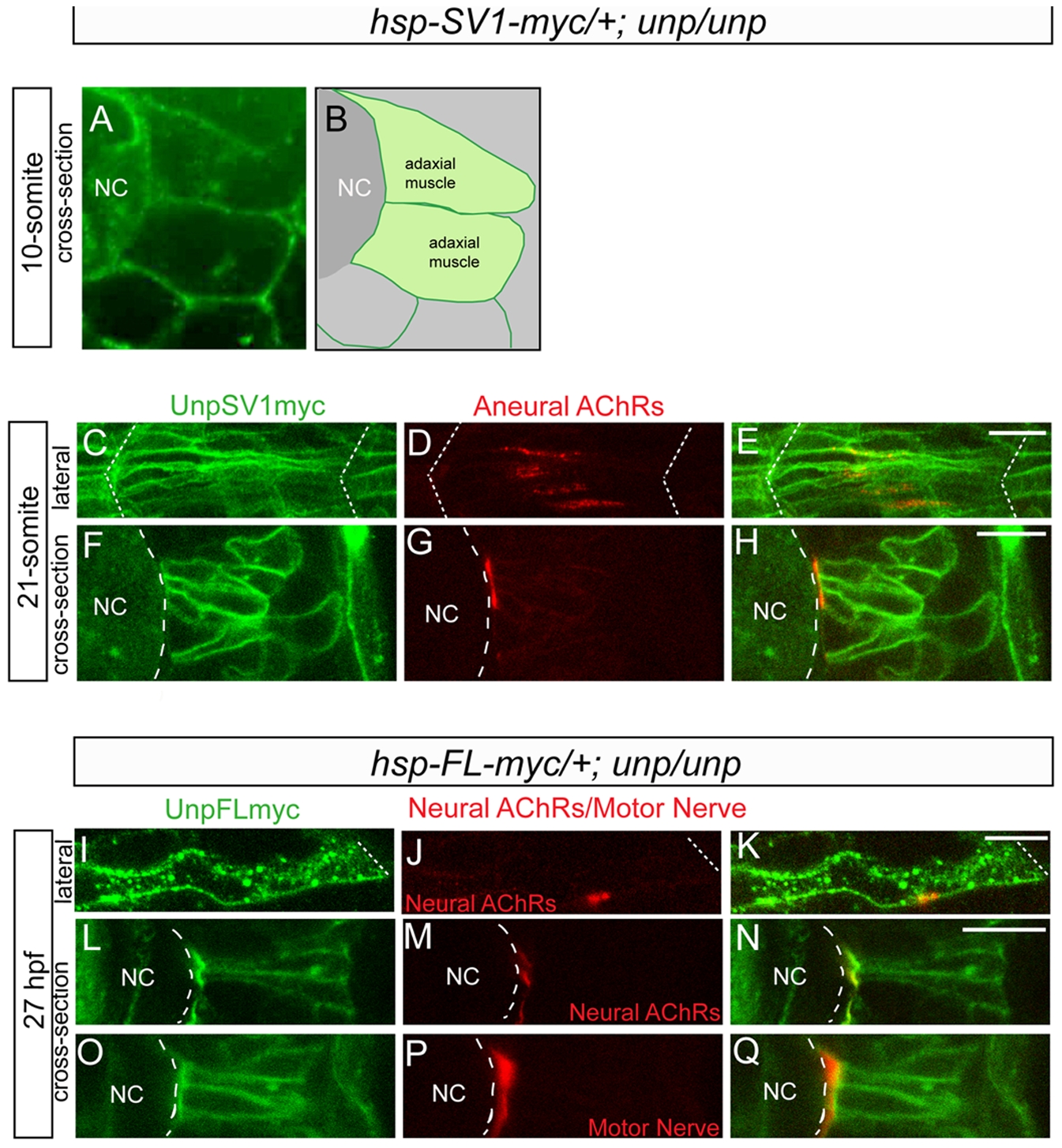
Fig. 6 Cellular localization of exogenous unplugged/MuSK protein and AChR clusters.
(A) Tg(hsp70l:unplugged SV1-myc); unpluggedbr307/br307 embryos at the 10-somite stage after the HS treatment, stained for anti-Myc Ab (green). Prior to the appearance of AChR clusters Unplugged SV1 protein is distributed evenly on the surface of adaxial muscle cells. (B) Diagram of A. (C–H) Tg(hsp70l:unplugged SV1-myc); unpluggedbr307/br307 embryos at the 21-somite stage after the HS treatment, stained for anti-Myc Ab (green) and AChR clusters (α-BTX, red). Lateral views (C–E) and cross-sectional views (F–H) show that Unplugged SV1 protein is distributed throughout the surface of muscle cells, but prepatterned clusters are localized at the center and the medial side of the cell surface. (I–Q) Tg(hsp70l:unplugged FL-myc); unpluggedbr307/br307 embryos at 27 hpf after HS treatment, stained for anti-Myc Ab (green, I–Q) and AChR clusters (α-BTX, red, J,K,M,N) or znp1(red, P–Q). Unplugged FL protein is expressed throughout the cell membrane as shown in lateral views (I–K), or cross-sectional views, (L–Q). Neural AChR clusters and motor axons are located at the center and the medial side of the cell. Non-migratory adaxial cells at the horizontal myoseptum were imaged. White dashed lines mark the boundaries of the somite in (C–E, I–K) or indicate the position of notochord in (A, B, F–H, L–Q). NC, notochord. Scale bars: 10 μM.