
|
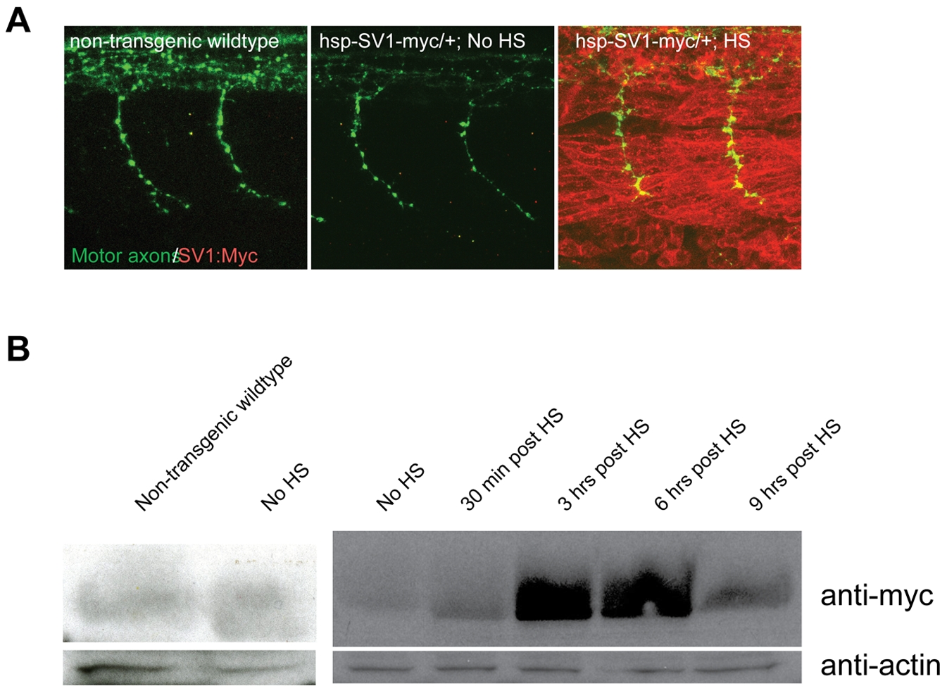
Fig. 2 Transgene expression after heat-shock (HS) induction.
(A) Lateral views of 26 hpf whole-mount embryos stained with anti znp1 (notor axons, green) and anti myc (red) antibodies. No Myc protein expression is detectable in non-transgenic wildtype and transgenic embryos, but is ubiquitously detectable 10 minutes after a 40-min HS treatment in transgenic embryos. HS treatment was performed at 24hpf for 40 minutes, embryos were fixed and processed at 26 hpf. (B) unplugged/MuSK-myc fusion protein was visualized by western blot with an anti-myc antibody (1:500) in non-transgenic wildtype, transgenic non-heat shocked controls, and 30 minutes, 3 hours, 6 hours and 9 hours after a 40-min HS induction of Tg(hsp70l:unplugged SV1-myc). An anti-actin antibody (1:500) is used as a loading control. Tg(hsp70l:unplugged FL-myc) shows similar results (data not shown). HS treatment was performed at 24hpf for 40 minutes, embryos were fixed and processed at indicated times post heat shock.