
|
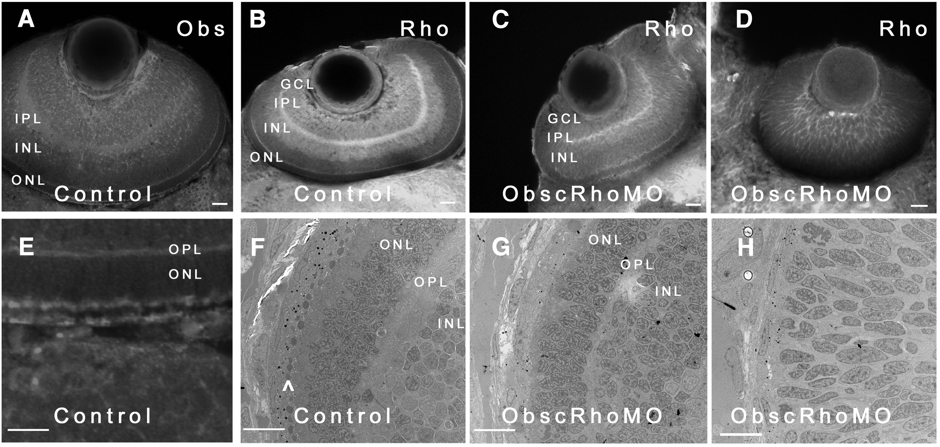
Fig. 9 Eye phenotype of obscurin A RhoGEF embryos. Control (A, B, E) and obscurin RhoGEF (C, D) morphant embryos were immunolabeled for Obscurin (A,E) or RhoA (B–D) at 72 hpf. In the control embryo, distinct retinal layers are noted [ganglion cell layer (GCL), inner plexiform layer (IPL), inner nuclear layer (INL), outer plexiform layer (OPL), and outer nuclear layer (ONL)]. Obscurin is expressed in each of the layers, usually at the cell periphery with the greatest abundance in the axon-rich IPL (A) and at the apical and basal aspects of the ONL (E) in a distribution similar to RhoA (A, B). In the mildly affected obscurin A RhoGEF morphant embryos (C), rudimentary GCL, IPL, INL, OPL, and ONL are noted while the more severely affected embryos do not develop identifiable layers (D). On ultrastructural analysis, there is significant pigment deposition and a well developed photoreceptor layer (^) in the retina of a control embryo (F) while even a mildly affected morphant embryo (G) has very little pigmentation and no identifiable photoreceptors. A more severely affected embryo (H) lacks even rudimentary layers and the cells remain elongated and poorly differentiated. Scale bars are 20 μm (A–D) and 10 μm (E–H).
Reprinted from Developmental Biology, 337(2), Raeker, M.O., Bieniek, A.N., Ryan, A.S., Tsai, H.J., Zahn, K.M., and Russell, M.W., Targeted deletion of the zebrafish obscurin A RhoGEF domain affects heart, skeletal muscle and brain development, 432-443, Copyright (2010) with permission from Elsevier. Full text @ Dev. Biol.