
|
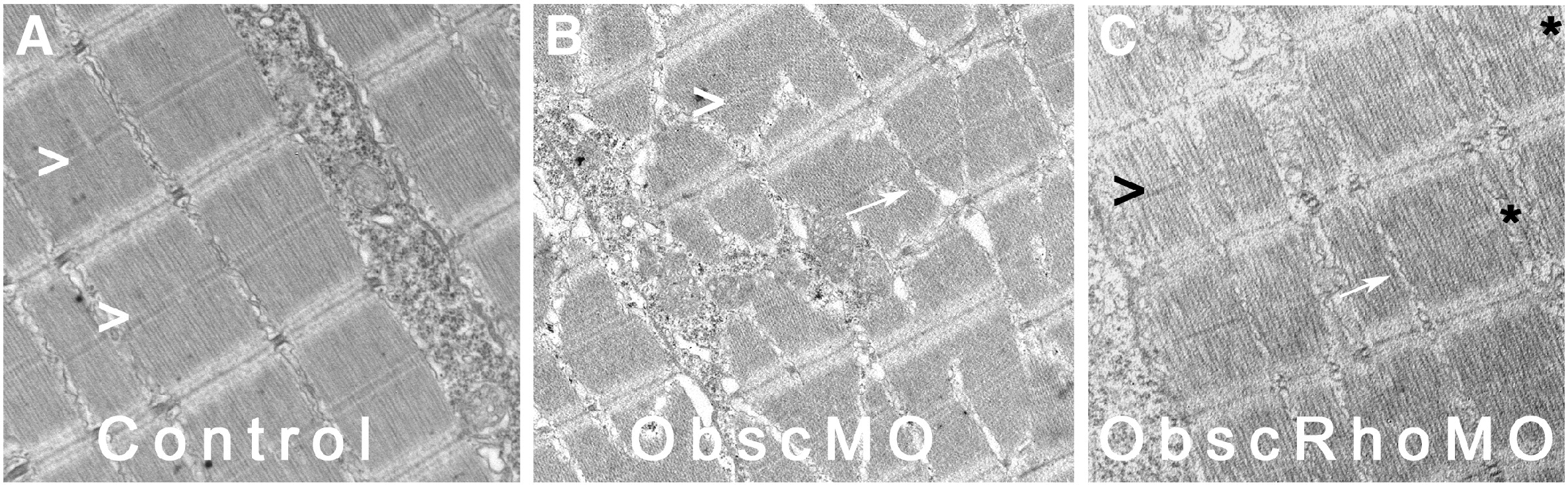
Fig. 5 Absence of consistent M bands in obscurin A RhoGEF skeletal muscle at 72 hpf. Control embryos (A) consistently demonstrate well-formed M bands (>) in skeletal myofibrils by 72 hpf. Obscurin A morphant embryos (B), although they display marked abnormalities in myofibrillar architecture, also consistently demonstrate mature M bands (>). In contrast, skeletal myofibrils in obscurin A RhoGEF morphant embryos (C) demonstrated occasional absence of M bands (∗) in otherwise mature-appearing myofibrils. Note that the extrajunctional SR in the ObscMO embryo is markedly disorganized (B: arrow), but is much more normal appearing in the ObscRhoMO embryos (C: arrow).
Reprinted from Developmental Biology, 337(2), Raeker, M.O., Bieniek, A.N., Ryan, A.S., Tsai, H.J., Zahn, K.M., and Russell, M.W., Targeted deletion of the zebrafish obscurin A RhoGEF domain affects heart, skeletal muscle and brain development, 432-443, Copyright (2010) with permission from Elsevier. Full text @ Dev. Biol.