
|
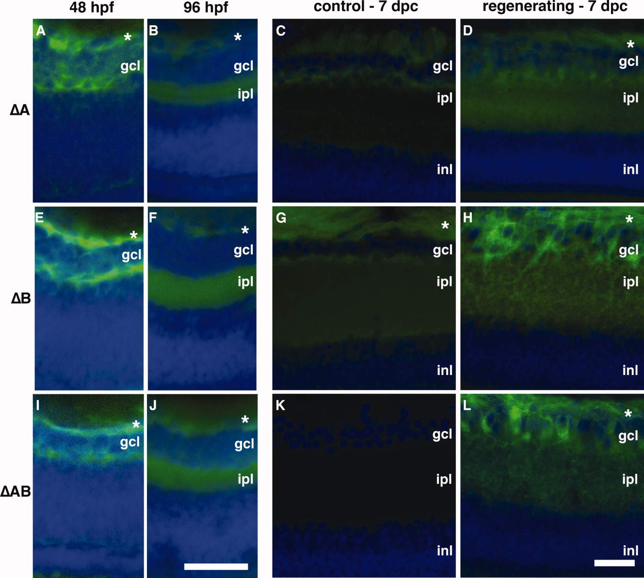
Fig. 5 Transgene expression in regenerating retina requires region C. Native GFP expression (green) and DAPI staining (blue) in transverse sections of embryonic (48 hpf; A, E, I), larval (4 dpf; B, F, J), and adult retinas (control; C, G, K; and regenerating; D, H, L) from promoter deletion GFP reporter lines: ΔA (A-D); ΔB (E-H); ΔAB (I-L). All lines displayed the same spatial and temporal pattern of expression in the developing retina (A, B, E, F, I, J) as previously observed for the full-length promoter (see Fig. 2A, B). Unlike ΔABC reporter lines, which did not express GFP in regenerating adult retina (see Fig. 2G, H), addition of the C region in the ΔAB lines was sufficient to restore regenerative expression (L). (*) Axons of RGCs in fiber layer. dpc, days post-crush; gcl, ganglion cell layer; ipl, inner plexiform layer; inl, inner nuclear layer. Developing retina scale bar (J) = 25 μm. Adult retina scale bar (L) = 50 μm.