
Image
|
Figure Caption
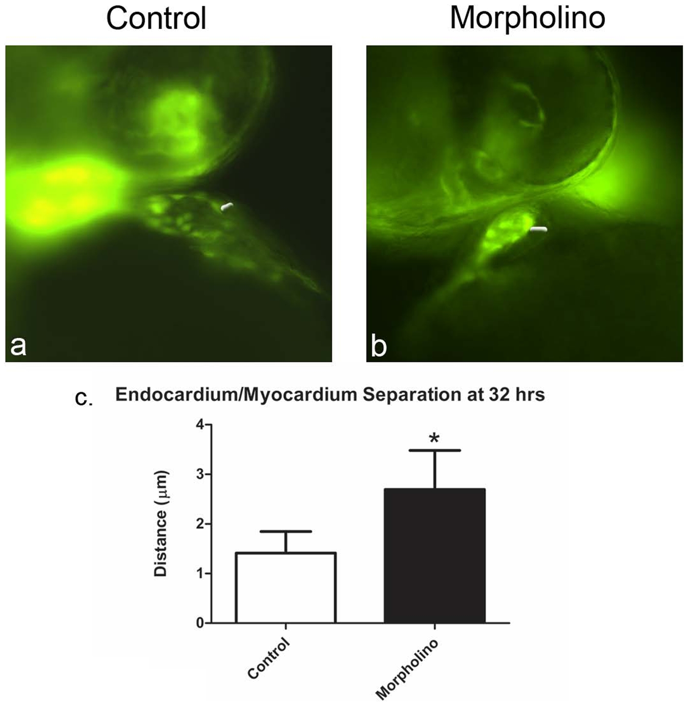
Fig. 6 VE-cadherin knockdown in fli1:GFP embryos reproduces and allows measurement of endocardial/myocardial separation at 32 hpf.
Images taken from movies of transgenic fli:GFP embryos that were injected with either control MO or VE-cad MO and observed with fluorescence microscopy (a,b). These images demonstrate increased endocardial/myocardial separation. The average measured distance between endocardium and myocardium (white arrows) differed significantly between the two groups, with the knockdown embryos consistently having a larger separation between the two layers (c).
Figure Data
Acknowledgments
This image is the copyrighted work of the attributed author or publisher, and
ZFIN has permission only to display this image to its users.
Additional permissions should be obtained from the applicable author or publisher of the image.
Full text @ PLoS One