
|
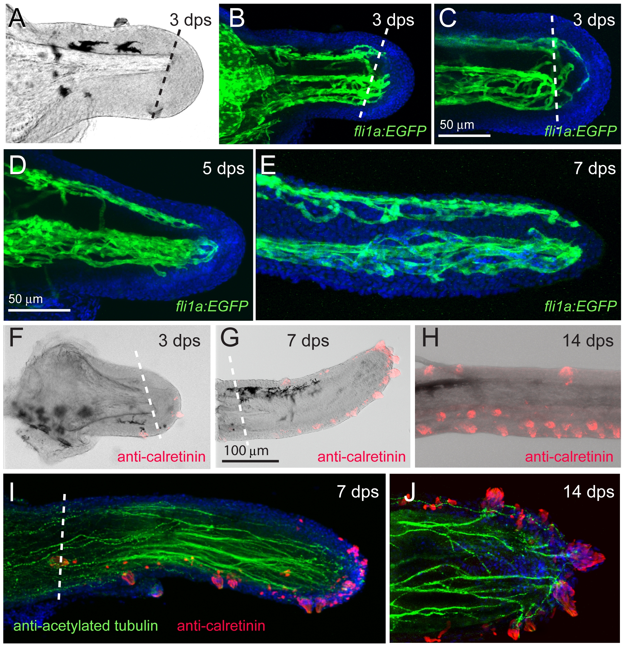
Fig. 10 Regeneration of the barbel vasculature, taste buds, and sensory nerves.
A) Transmitted light image of a Tg(fli1a:EGFP) maxillary barbel stump 3 days post surgery (dps). The distal end projects right. B) Confocal image of the vasculature in A. Endothelial sprouts (green) project distally past the plane of section and appear to bridge the dorsal (top) and ventral (bottom) vessels. Nuclei are counterstained blue (DAPI). C) Confocal reconstruction of a second regenerating barbel (3 dps). Similar sprouting is visible, as well as several isolated endothelial cells migrating underneath the wound epithelium. D–E) Regeneration of the vasculature at 5 and 7 days post surgery. Two streams of endothelial cells are visible; a dorsal stream (top) and a ventral stream (bottom). Both sets of vessels are more torturous than those of the original barbel (e.g., Fig. 4E). F–H) Regeneration of the taste buds 3–14 days post surgery (dps). Calretinin-positive cells appear at the tip within 3 days (F). By 7 dps, the distribution of taste buds on the ventral side and distal tip resembles the normal adult pattern (G). The ventral taste buds of a 14-day regenerate (H) are arranged in a typical double row (compare to Fig. 6D). I, J) Regenerating maxillary barbels are densely innervated with long axons (anti-acetylated tubulin, green) projecting to the bases of the taste buds (anti-calretinin, red).