
|
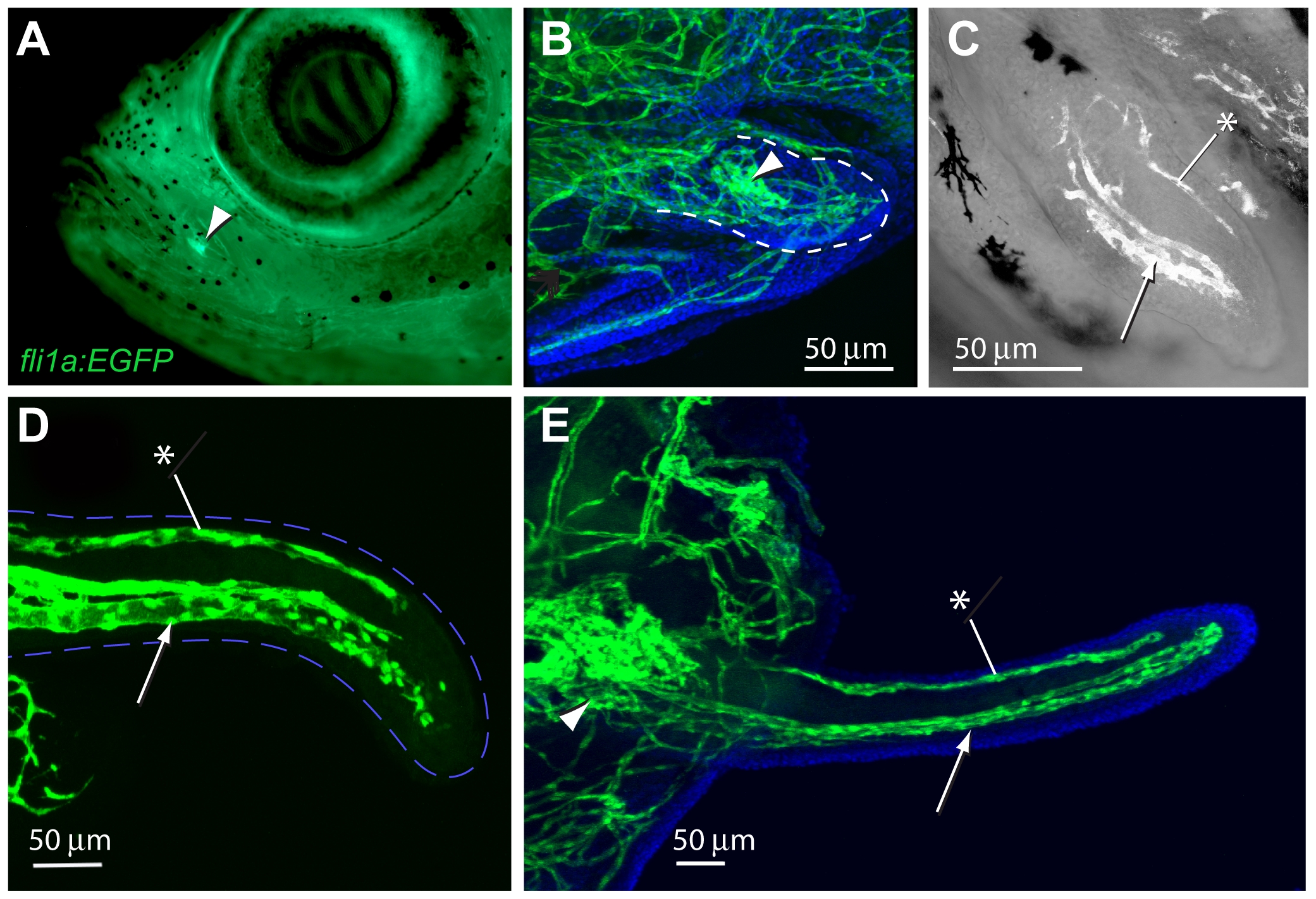
Fig. 4 Development of the maxillary barbel vasculature in Tg(fli1a:EGFP) transgenic zebrafish.
A) In vivo image of a juvenile Tg(fli1a:EGFP) zebrafish (10–12 mm standard length), in which all endothelial cells fluoresce green. The base of the future maxillary barbel is visible externally as a bright green confluence of blood vessels on the posterior ventral corner of the maxilla (arrowhead). B) 75 µm barbel. Confocal reconstruction of the early barbel bud circulation; anterior is to the left. A confluence of green vessels is visible at the base of the barbel (arrowhead). Smaller endothelial sprouts invade the bud proper (within the dotted line). Nuclei are counterstained blue (DAPI). C) 125 μm barbel. Two streams of endothelial cells are visible; a larger ventral stream (arrow), which will form the capillary loop, and a smaller dorsal stream (asterisk), which will form the putative lymphatic. In this focal plane, the proximal plexus of vessels is not visible. D) 300 μm barbel. The proximal ends of the ventral and dorsal vessels appear patent and lined with flattened endothelial cells. The distal ends of the vessels are composed of loose amoeboid cells with filipodia projecting into the surrounding tissue. The outline of the barbel is dashed blue. E) 600 μm barbel. The circulation at this stage consists of a closed capillary loop ventrally and a single, blind-end vessel dorsally. The proximal vascular plexus is greatly enlarged. Nuclei are counterstained blue (DAPI). Arrowhead = proximal vascular plexus; arrow = ventral vessels; asterisk = dorsal vessel (putative lymphatic).