
|
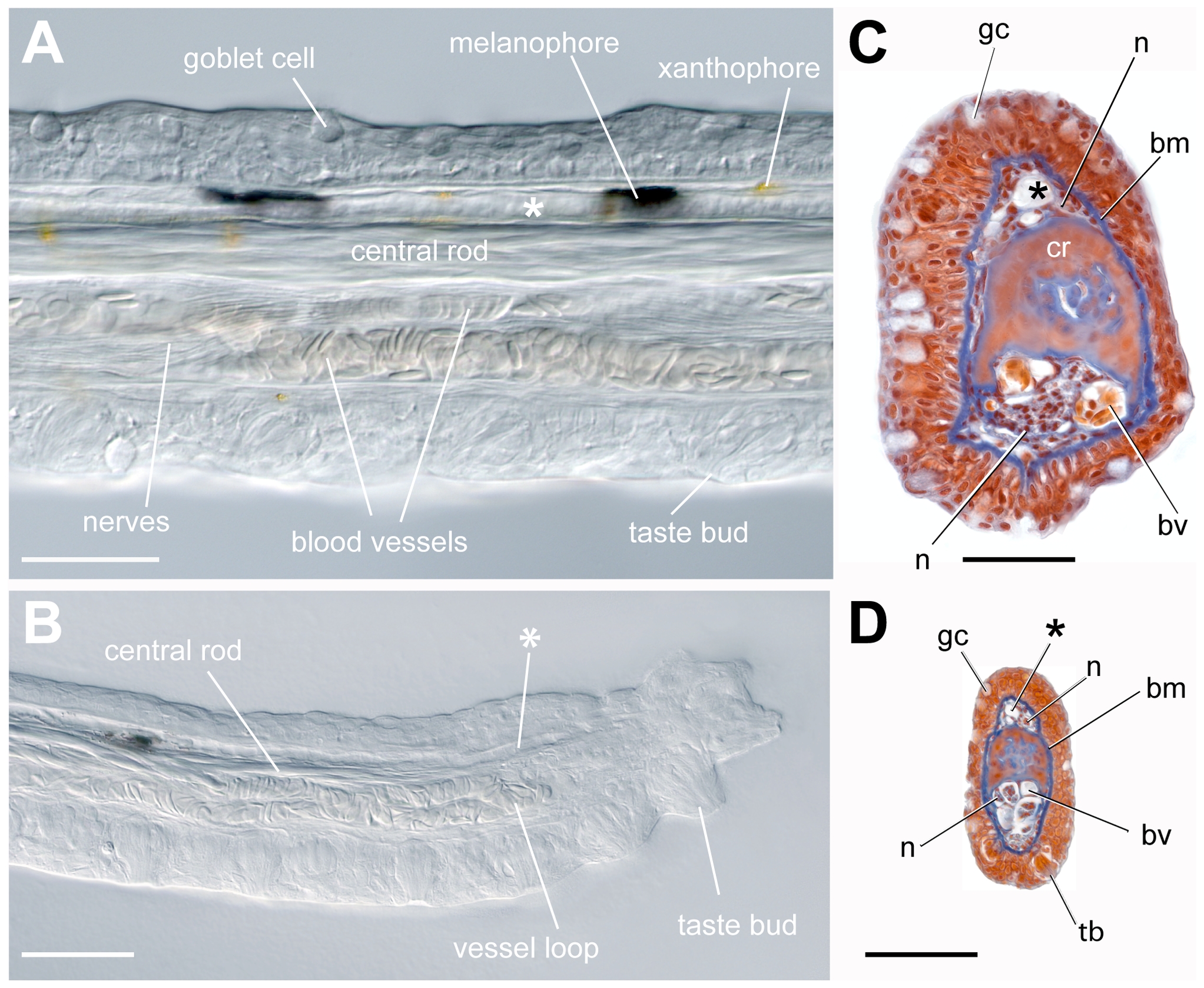
Fig. 2 Whole-mount and sectional views of the adult maxillary barbel.
A) Differential interference contrast (DIC) image of an adult maxillary barbel shaft fixed and cleared in 50% glycerol. All of the central tissues are visible through the transparent outer epithelium. A putative lymph vessel (*) lies dorsal to the central rod. All scale bars = 100 μm. B) DIC image of an adult maxillary barbel tip at the same scale as A. The central rod is reduced to a narrow band of fibers. The ventral vasculature terminates in a capillary loop packed with erythrocytes, while the lymphatic (*) terminates in a blind, tapered end. The ventral epithelium and distal tip carry numerous taste buds. C) Representative cross-section of an adult maxillary barbel at the level shown in A. Nuclei are dark red, erythrocytes orange, and basal laminae/connective tissues blue (Mallory′s trichrome). D) Representative cross-section at the level shown in B. bm = basement membrane; bv = blood vessel; gc = goblet cell; n = nerve fibers; tb = taste bud; * = putative lymph vessel (for explanation see text).