
|
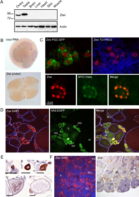
Fig. 1 Ziwi Subcellular Localization
(A) Western blot shows specific expression of Ziwi in testis and ovary.
(B) Ziwi protein is maternally provided and localizes to granules at the cleavage planes in four cell embryos similar to vasa mRNA.
(C) At 24 hpf Ziwi (red) localizes to distinct perinuclear granules in the PGCs (upper two panels). These granules also contain Vasa (lower three panels).
(D) Ziwi (red) in ovary is expressed in oogonia and stage I oocytes. Vasa (in green) is expressed in all stages of oogenesis. Stages of oogenesis are oogonia (OG), stage I oocytes (7–140 µm; I), stage II oocytes (140–340 μm; II), stage III oocytes (340–690 μm; not in picture; III), and stage IV oocytes (0.69–0.73 mm; Selman et al., 1993).
(E) Ziwi expression in various stages of oocytes is visualized by DAB staining to reveal lower levels of expression in later stage oocytes. I indicates oogonia; II indicates Stage I; III indicates Stage II; IV indicates Stage III; and V indicates Early stage V. Scale bar is 100 μm.
(F) Ziwi (left panel, red, or right panel, brown) in testis is expressed in spermatogonia and spermatocytes. Stages of spermatogenesis are spermatogonia (SG), spermatocytes (SC), spermatids (ST), and sperm (S).
Reprinted from Cell, 129(1), Houwing, S., Kamminga, L.M., Berezikov, E., Cronembold, D., Girard, A., van den Elst, H., Filippov, D.V., Blaser, H., Raz, E., Moens, C.B., Plasterk, R.H., Hannon, G.J., Draper, B.W., and Ketting, R.F., A role for Piwi and piRNAs in germ cell maintenance and transposon silencing in Zebrafish, 69-82, Copyright (2007) with permission from Elsevier. Full text @ Cell