
Image
|
Figure Caption
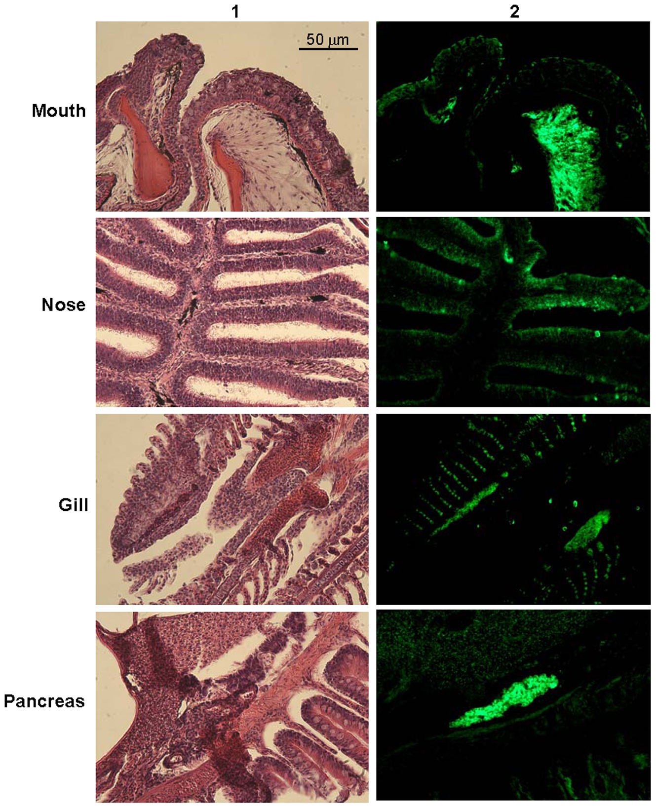
Fig. 2 Immunohistochemistry of trypsin in zebrafish.
Zebrafish were dissected and sections of mouth, nose, gill, and pancreas were stained with H&E (1) and subjected to immunohistochemistry analysis using polyclonal rabbit antisera against human trypsin (PRAHT) (2). Alexa goat anti-rabbit IgG 488 antisera were used as the secondary antibody. Non-immunized rabbit IgG was used in control experiments (images not shown). 20X lens was used.
Figure Data
Acknowledgments
This image is the copyrighted work of the attributed author or publisher, and
ZFIN has permission only to display this image to its users.
Additional permissions should be obtained from the applicable author or publisher of the image.
Full text @ PLoS One