
Image
|
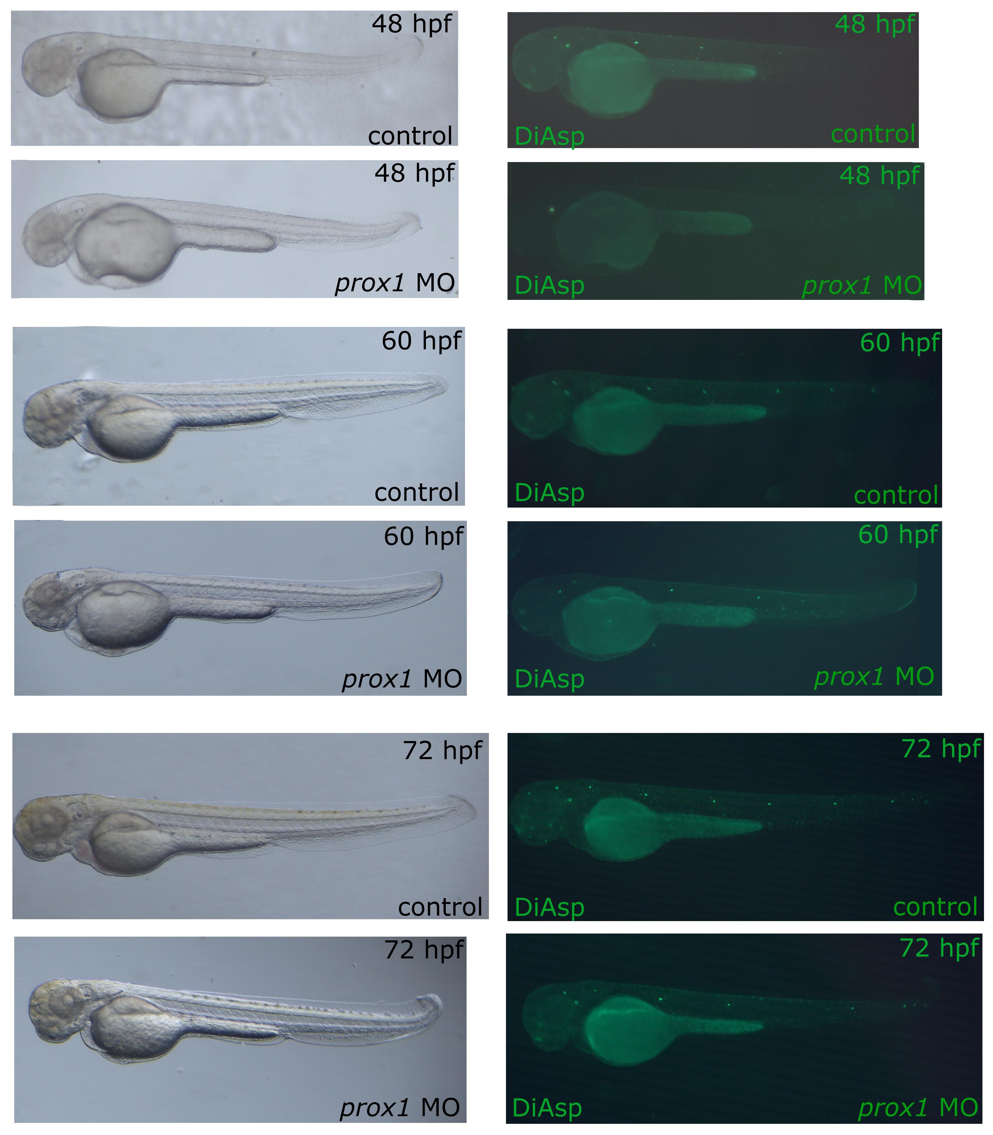
Figure Caption
Fig. S2 DiAsp staining in control and prox1 loss of function embryos at 72 hpf. As at 48 hpf, also at 60 and 72 hpf, prox1 MO injected embryos still presented a decrease number of DiAsp positive cells in neuromasts in comparison to control embryos at the same developmental stage, indicating that the effect is not due to developmental delay of morphant embryos.
Acknowledgments
This image is the copyrighted work of the attributed author or publisher, and
ZFIN has permission only to display this image to its users.
Additional permissions should be obtained from the applicable author or publisher of the image.
Full text @ BMC Dev. Biol.