
|
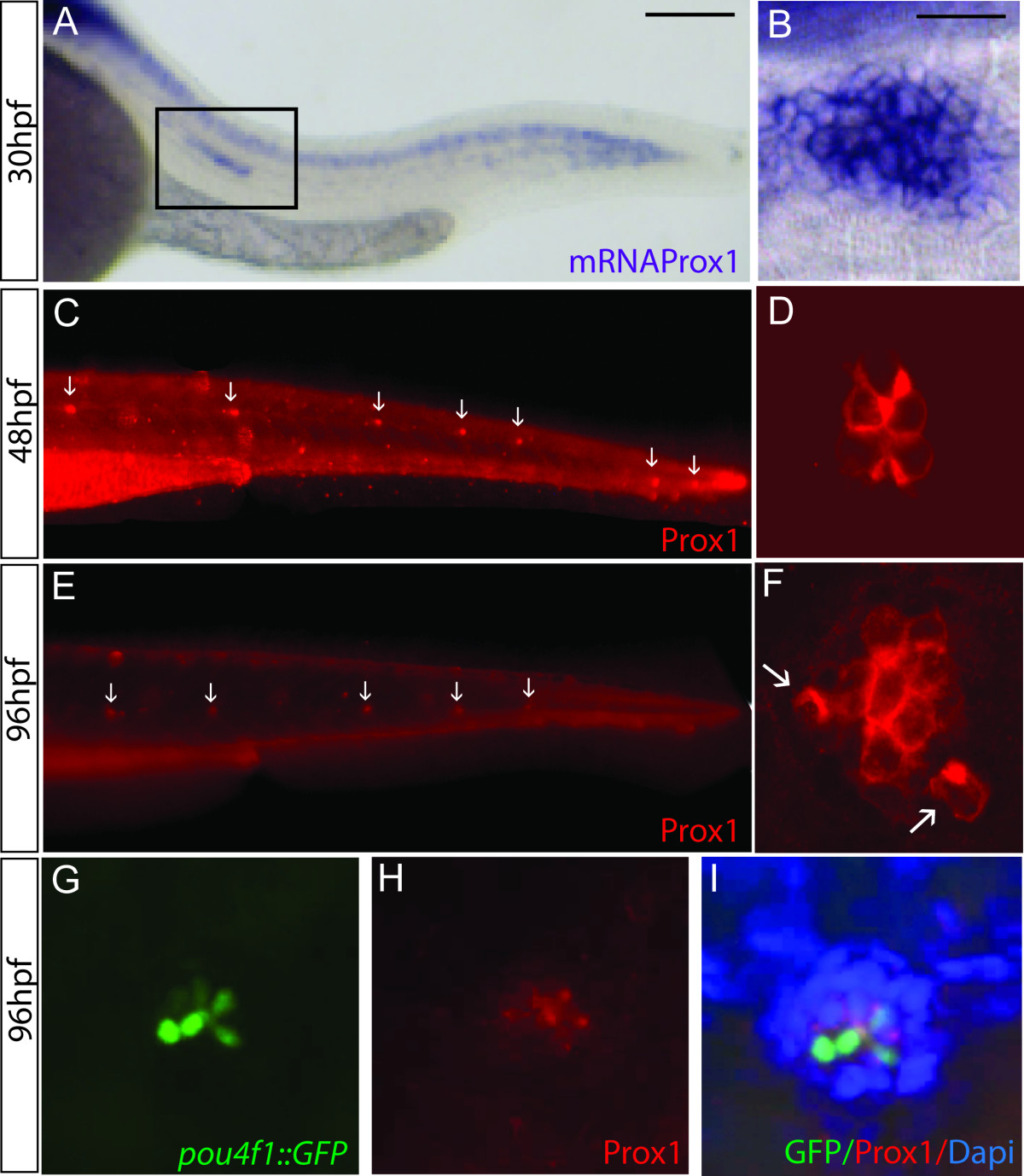
Fig. 1 Prox1 expression in the lateral line system of zebrafish embryos. (A) In situ hybridization of prox1 at 30 hpf shows expression in the CNS and in the lateral line migrating primordium (box). (B) Enlarged view of a prox1 positive deposited neuromast in the posterior lateral line at 30 hpf. (C, E) Immunofluorescence using an anti-Prox1 antibody at 48 hpf and 96 hpf, arrows indicate the deposited neuromasts. (D, F) Close up of Prox1 expression in a neuromast at the two stages examined. (G, H, I) Immunofluorescence labeling in neuromasts with anti GFP (G), anti Prox1 (H) and the cell nuclei with DAPI (I) in 96 hpf pou4f1::GFP transgenic larvae. Scale bar = 10 micron.