
|
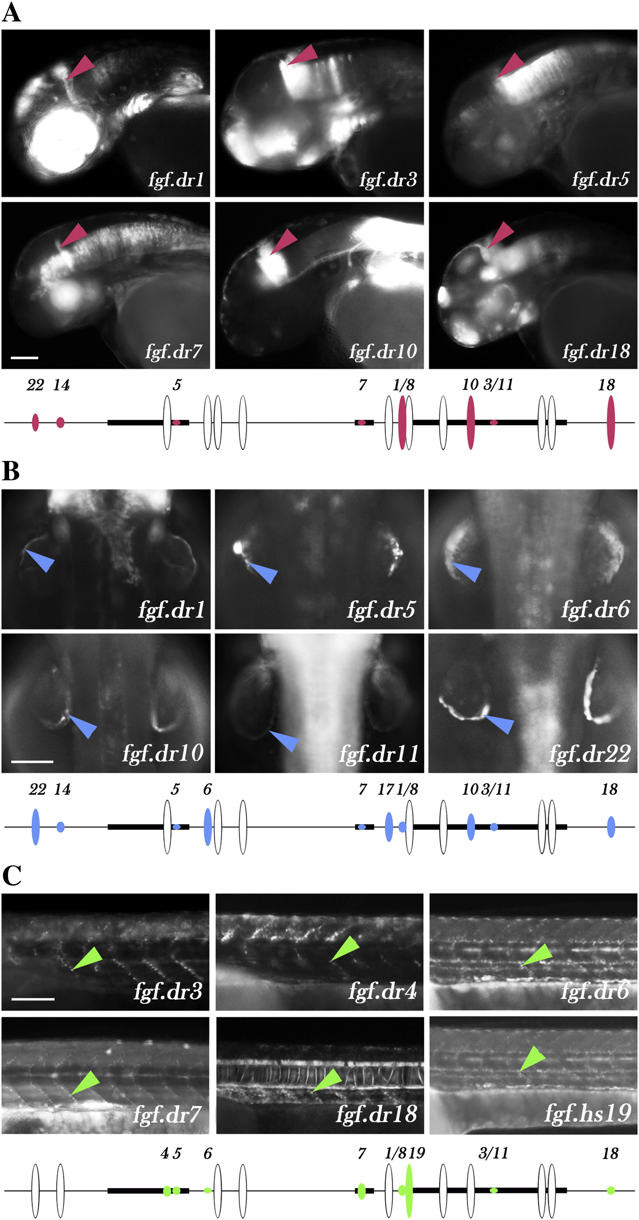
Fig. 6 Multiple transgenes with reporter expression in signaling centers. Certain structures (A: midbrain–hindbrain boundary; B: apical ectodermal ridge; C: anterior myotomes) that fall into the expression domain of fgf8a are regulated through a set of tested fgf8a elements. Upper panel shows examples of transgenic lines from regulatory elements that directed reporter protein expression to specific structures. Lower section shows all regulatory elements that are active in particular structures. The height of ovals indicates the percentage of transgenic lines from each element that drove GFP expression in a particular structure (white oval height equals 100%). The tetrapod-specific element fgf.hs19 is indicated in the place corresponding to the human locus. (A) Midbrain–hindbrain boundary (MHB) expression is regulated by 8 elements (pink) dispersed around fgf8a: fgf.dr1/8, fgf.dr3/11, fgf.dr5, fgf.dr7, fgf.dr10, fgf.dr14, fgf.dr18 and fgf.dr22. (B) Apical ectodermal ridge (AER) expression is regulated by 10 elements (blue): fgf.dr1/8, fgf.dr3/11, fgf.dr4, fgf.dr6, fgf.dr7, fgf.dr10, fgf.dr14, fgf.dr17, fgf.dr18 and fgf.dr22. (C) Reporter expression to anterior myotome and tail tip is directed by 8 elements (green): fgf.dr1/8, fgf.dr3/11, fgf.dr4, fgf.dr5, fgf.dr6, fgf.dr7, fgf.dr18 and fgf.hs19. Scale bar = 100 μm.
Reprinted from Developmental Biology, 336(2), Komisarczuk, A.Z., Kawakami, K., and Becker, T.S., Cis-regulation and chromosomal rearrangement of the fgf8 locus after the Teleost/tetrapod split, 301-312, Copyright (2009) with permission from Elsevier. Full text @ Dev. Biol.