
Image
|
Figure Caption
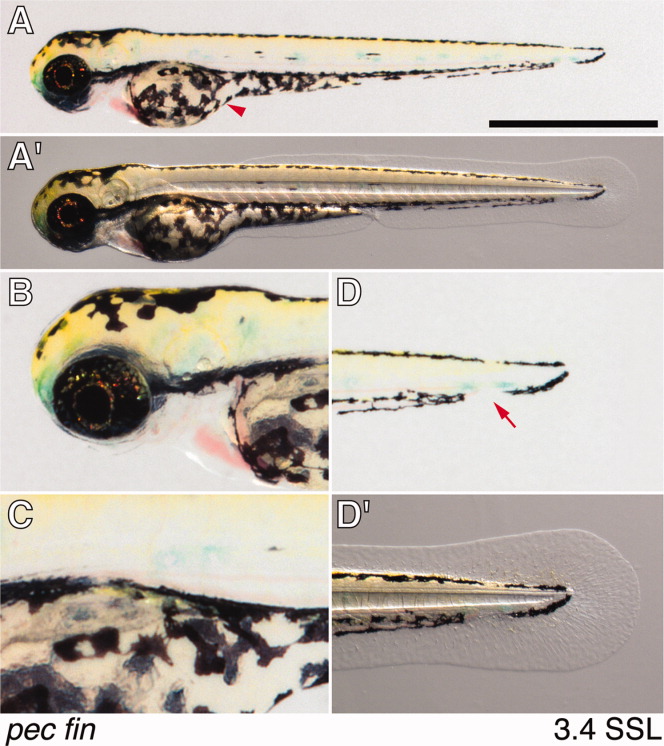
Fig. 32 Embryonic pec fin stage of Kimmel et al. ([1995]); 3.4 mm SL (standard length). A,A′: Whole body. Arrowhead, melanophores covering yolk ball. Scale bar = 1 mm. B: Head. C: Swim bladder, not yet inflated. C′: Tail. Arrow, gap in melanonophore pattern corresponding to region of caudal fin condensation.
Acknowledgments
This image is the copyrighted work of the attributed author or publisher, and
ZFIN has permission only to display this image to its users.
Additional permissions should be obtained from the applicable author or publisher of the image.
Full text @ Dev. Dyn.