
|
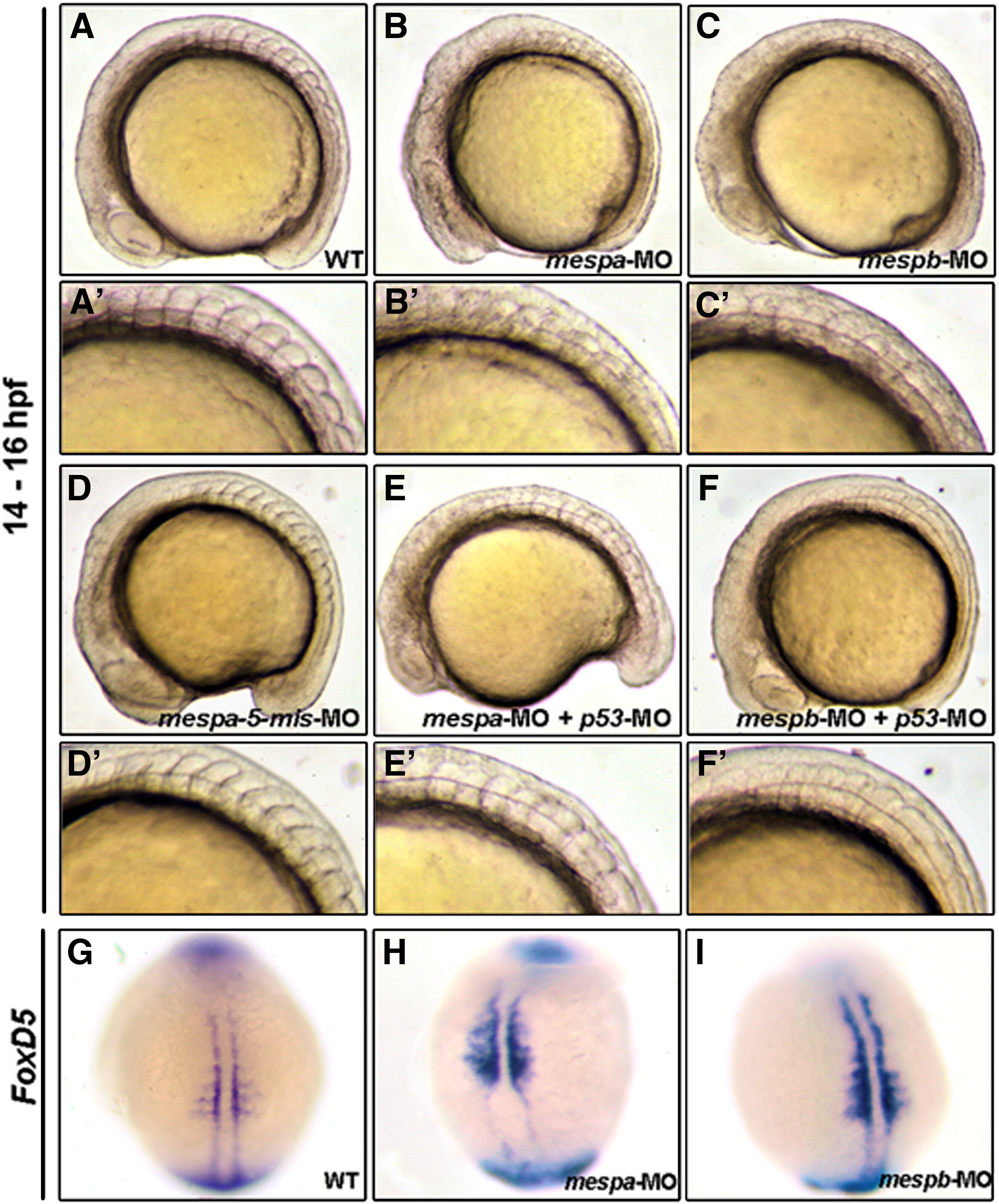
Fig. S6 Knockdown of either mespa or mespb disrupted somite formation, but the expression of FoxD5 appeared unaffected. (L-F) Lateral views of trunk somites in wild-type embryos (WT) and embryos injected with different materials as indicated. Panels A′-F′ were magnified, and the somite morphology is shown in panels A-F, respectively. Zebrafish WT embryos exhibited normally developed somites at 18-hpf (A and A′). The mespa-MO- (B, B′) and mespb-MO-injected (C and C′) embryos displayed defects in somite formation. Injection of a control MO, mespa-5mis-MO, which contains 5-bp, which are mismatched to the corresponding mespa-MO, resulted in a normal phenotype (D and D′). When embryos were co-injected with either mespa-MO combined with p53-MO (E and E′) or mespb-MO combined with p53-MO (F and F′), the somite structure was still disorganized. Using whole-mount in situ hybridization to detect the expression of FoxD5 in WT (G), mespa (H), and mespb (I) morphants at 12-hpf as indicated. Like WT control embryos (G), the striped pattern of FoxD5 in the anterior PSM was clearly detected in both mespa (H) and mespb (I) morphants.
Reprinted from Developmental Biology, 336(2), Lee, H.C., Tseng, W.A., Lo, F.Y., Liu, T.M., and Tsai, H.J., FoxD5 mediates anterior-posterior polarity through upstream modulator Fgf signaling during zebrafish somitogenesis, 232-245, Copyright (2009) with permission from Elsevier. Full text @ Dev. Biol.