
|
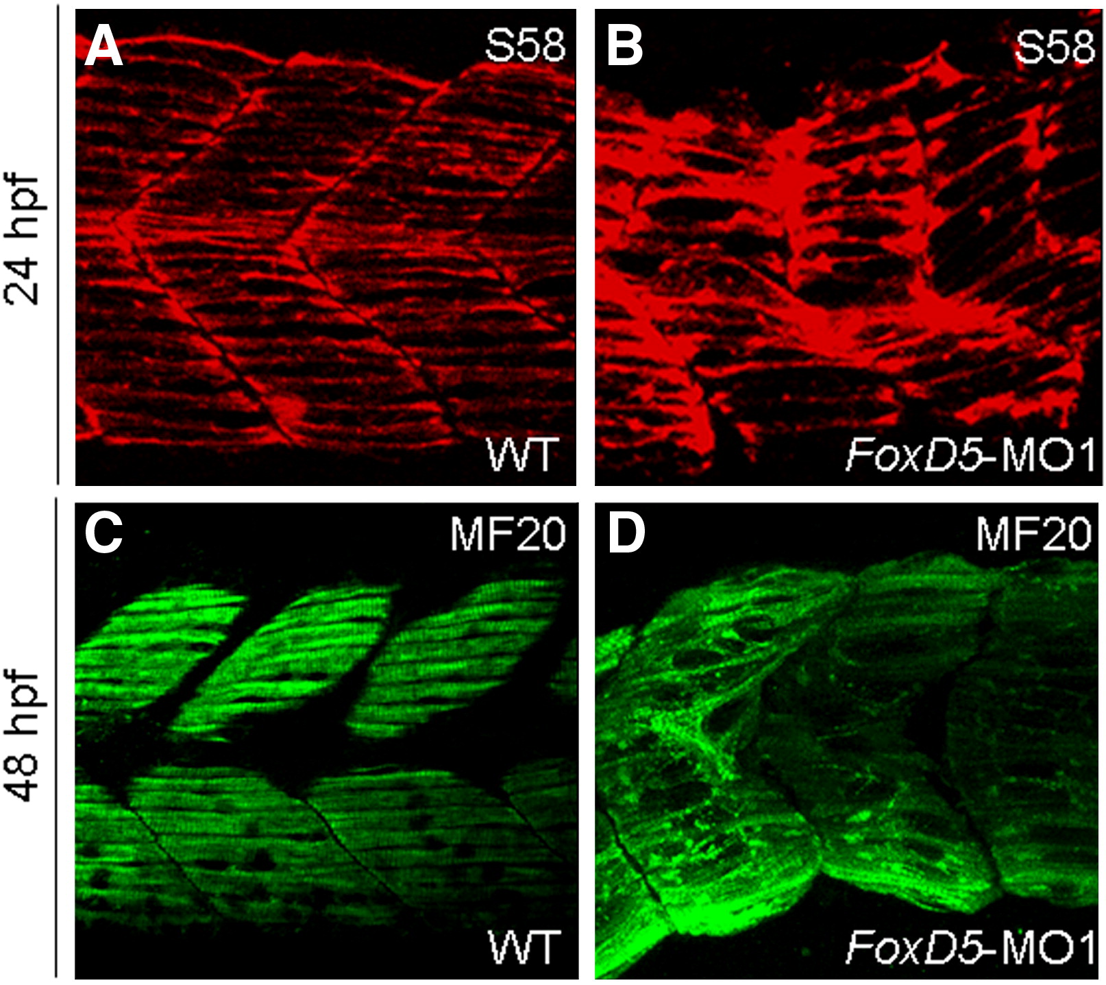
Fig. S5 Knockdown of FoxD5 caused the misalignment of myofibrils. The wild-type (WT) and FoxD5-MO-injected embryos at 24- and 48-hpf were collected and subjected to immunochemical staining with antibodies of S58 (Red) and MF20 (green). At 24 hpf, WT embryos exhibited a normal morphology of slow muscles, which revealed a clear arrangement of slow fibers (A). On the other hand, the FoxD5 morphants showed an irregular arrangement of slow muscle fibers (B). At 48 hpf, all muscles were stained by MF20. Normal alignment of myofibrils was observed in WT embryos (C), but misalignment of myofibrils was observed in FoxD5 morphants (D).
Reprinted from Developmental Biology, 336(2), Lee, H.C., Tseng, W.A., Lo, F.Y., Liu, T.M., and Tsai, H.J., FoxD5 mediates anterior-posterior polarity through upstream modulator Fgf signaling during zebrafish somitogenesis, 232-245, Copyright (2009) with permission from Elsevier. Full text @ Dev. Biol.