
|
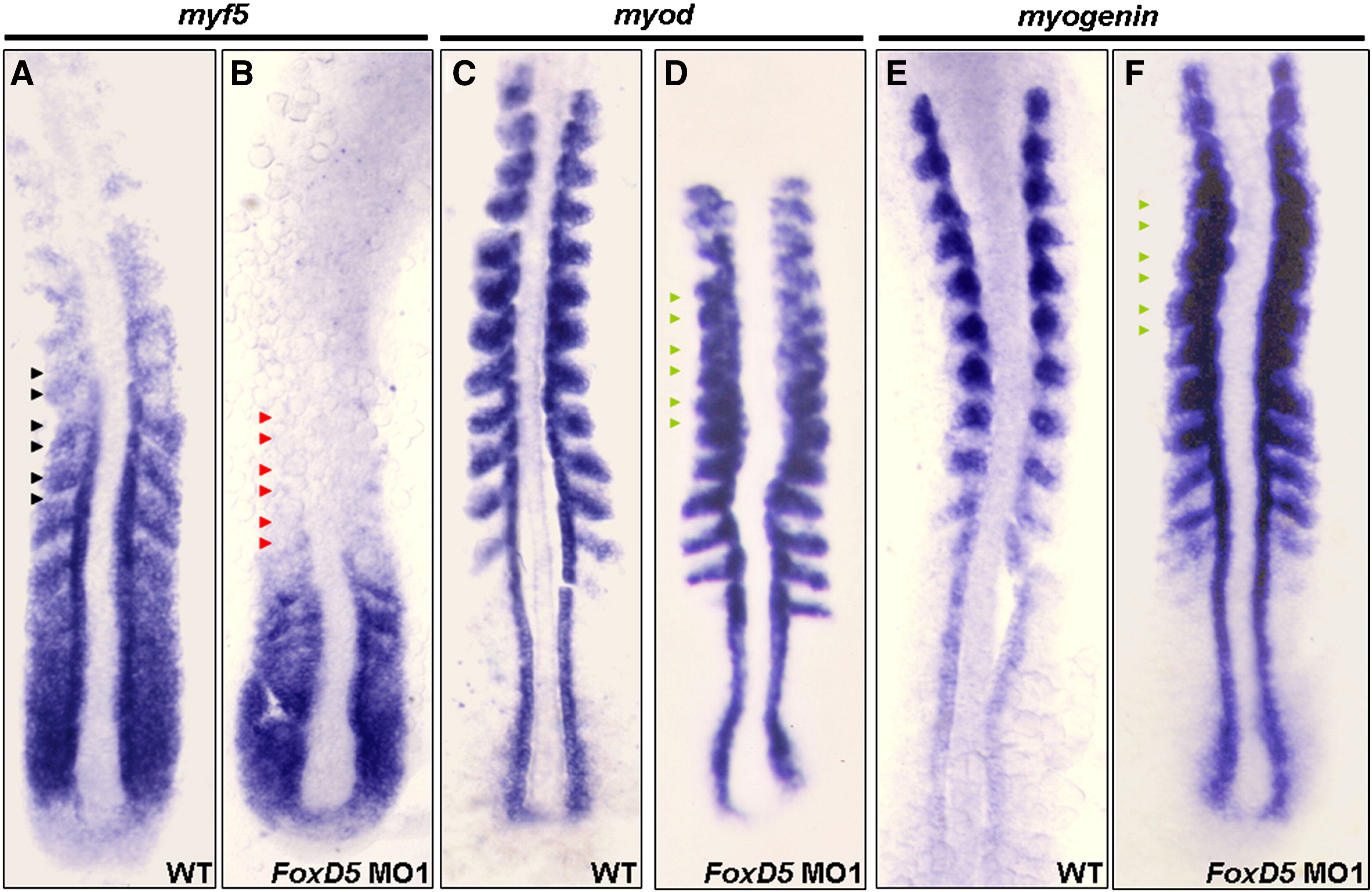
Fig. S4 Knockdown of FoxD5 reduced myf5 expression, but enhanced myod expression during somitogenesis. The wild-type embryos (WT) and FoxD5-MO-injected embryos at 14-hpf were collected, and WISH was performed with a probe of myf5 (A, B), myod (C, D), and myogenin (E, F). In WT (A), myf5 was expressed in the PSM in a gradient manner from faint intensity at anterior to strong intensity at posterior. In FoxD5 morphants (B), myf5 expression was reduced (red arrowhead), similar to that of WT in the PSM region. However, myf5 was not detected in the somites, as indicated by red arrowhead. In WT (C), myod was expressed at adaxial cells and the posterior part of a somite, but not in somite S-I, as indicated by blue arrowhead. In FoxD5 morphants (D), the myod expression pattern did not change, but the signal intensity was slightly up-regulated. In WT (E), myogenin was expressed at adaxial cells and the forming somites, including those from SI. In FoxD5 morphants (F), myogenin expression was no different from that of WT.
Reprinted from Developmental Biology, 336(2), Lee, H.C., Tseng, W.A., Lo, F.Y., Liu, T.M., and Tsai, H.J., FoxD5 mediates anterior-posterior polarity through upstream modulator Fgf signaling during zebrafish somitogenesis, 232-245, Copyright (2009) with permission from Elsevier. Full text @ Dev. Biol.