
Image
|
Figure Caption
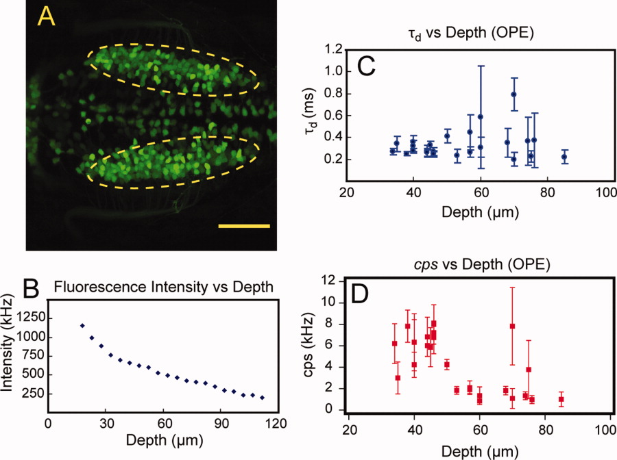
Fig. 2 Penetration depth study using one-photon excitation. A: Dorsal view of vagal motor neurons of 3 dpf embryo of Islet-1-EGFP transgenic line as indicated in the dashed ellipse. B: Fluorescence intensity measurements of EGFP-expressing motor neurons at different depths. C: Diffusion time measurements by fluorescence correlation spectroscopy (FCS) at different cell depths. D: cps of FCS measurements at different cell depths. Scale bar = 50 μm.
Acknowledgments
This image is the copyrighted work of the attributed author or publisher, and
ZFIN has permission only to display this image to its users.
Additional permissions should be obtained from the applicable author or publisher of the image.
Full text @ Dev. Dyn.