
Image
|
Figure Caption
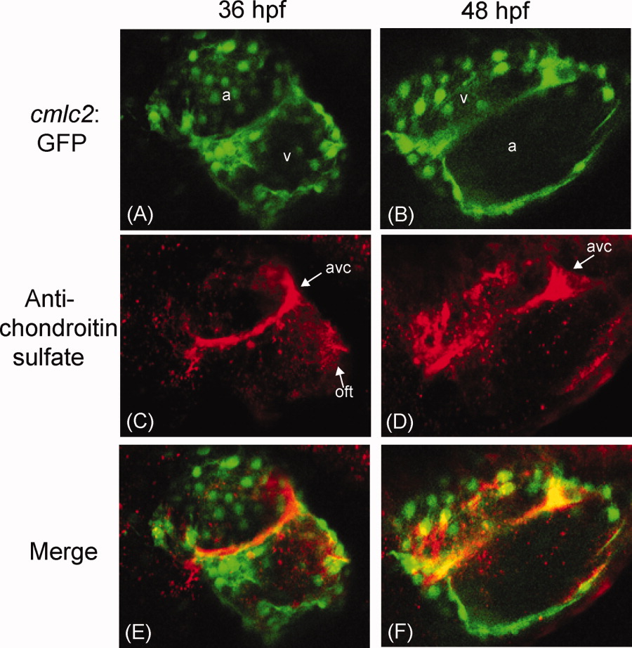
Fig. 2 Confocal images of Danio rerio hearts stained with an anti-chondroitin sulfate antibody at 36 and 48 hours postfertilization (hpf). A,B: Confocal microscopy of Danio rerio cis/trans-decahydro-2-napthol-β-D-xyloside (DX) containing the myocardium specific cmlc2:GFP marker (green). C,D: Confocal microscopy of the same Danio rerio images stained with a CS-specific antibody (red). E,F: Merged images. a, atrium; v, ventricle; avc, atrioventricular canal forming region; oft, outflow tract.
Figure Data
Acknowledgments
This image is the copyrighted work of the attributed author or publisher, and
ZFIN has permission only to display this image to its users.
Additional permissions should be obtained from the applicable author or publisher of the image.
Full text @ Dev. Dyn.