
|
Fig. S5
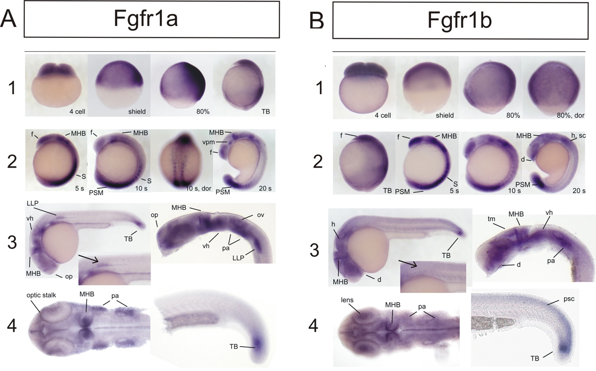
Expression of fgfr1a and fgfr1b during Embryogenesis of the Zebrafish
In situ hybridization at early stages of development with specific probes for fgfr1a (A) and fgfr1b (B). Both fgfr1a and fgfr1b are maternally provided and are ubiquitously expressed until midgastrulation (A 1 and B 1). At this stage the ventral expression starts disappearing. fgfr1b is also downregulated in dorsal midline and dorsal margin. From tail bud until mid-somitogenesis the ubiquitous dorsal expression of fgfr1a becomes restricted to the forebrain, the MHB, somites and presomitic mesoderm (A 2 and B 2). It is also found at low levels in the hindbrain. During later somitogenesis (20 somites – 24 hpf), fgfr1a transcript can be found in pharyngeal arches, the MHB, several domains of the telencephalon and the diencephalon, including the optic stalk and the olfactory placode (A 3, 4). fgfr1a is specifically expressed in the lateral line primordium. In the tail, expression persists in the posterior somites and tail bud, and then gets restricted to the tail bud by 24 hpf. In comparison, fgfr1b is expressed in broader domains of diencephalon, midbrain, cerebellum and ventral hindbrain (B 2, 3). The telencephalon is completely devoid of fgfr1b, which instead is expressed in the retina. There is a weak expression in the neural crest and inner cell layer of otic vesicles, whereas the expression in the trunk and somites seems to stay diffuse at low levels. Posterior somites and presomitic mesoderm express fgfr1b strongly until after 24 hpf. Abbreviations: d - diencephalon, f – forebrain, h – hindbrain, MHB – midbrainhindbrain boundary, LLP – lateral line primordium, op – olfactory placode, ov – otic vesicle, pa – pharyngeal arches, psc – posterior spinal cord, PSM – presomitic mesoderm, s – somites, sc – anterior spinal cord, TB – tail bud, tm – tectum of midbrain, vh – ventral hindbrain, vm – ventral posterior midbrain.