
Image
|
Figure Caption
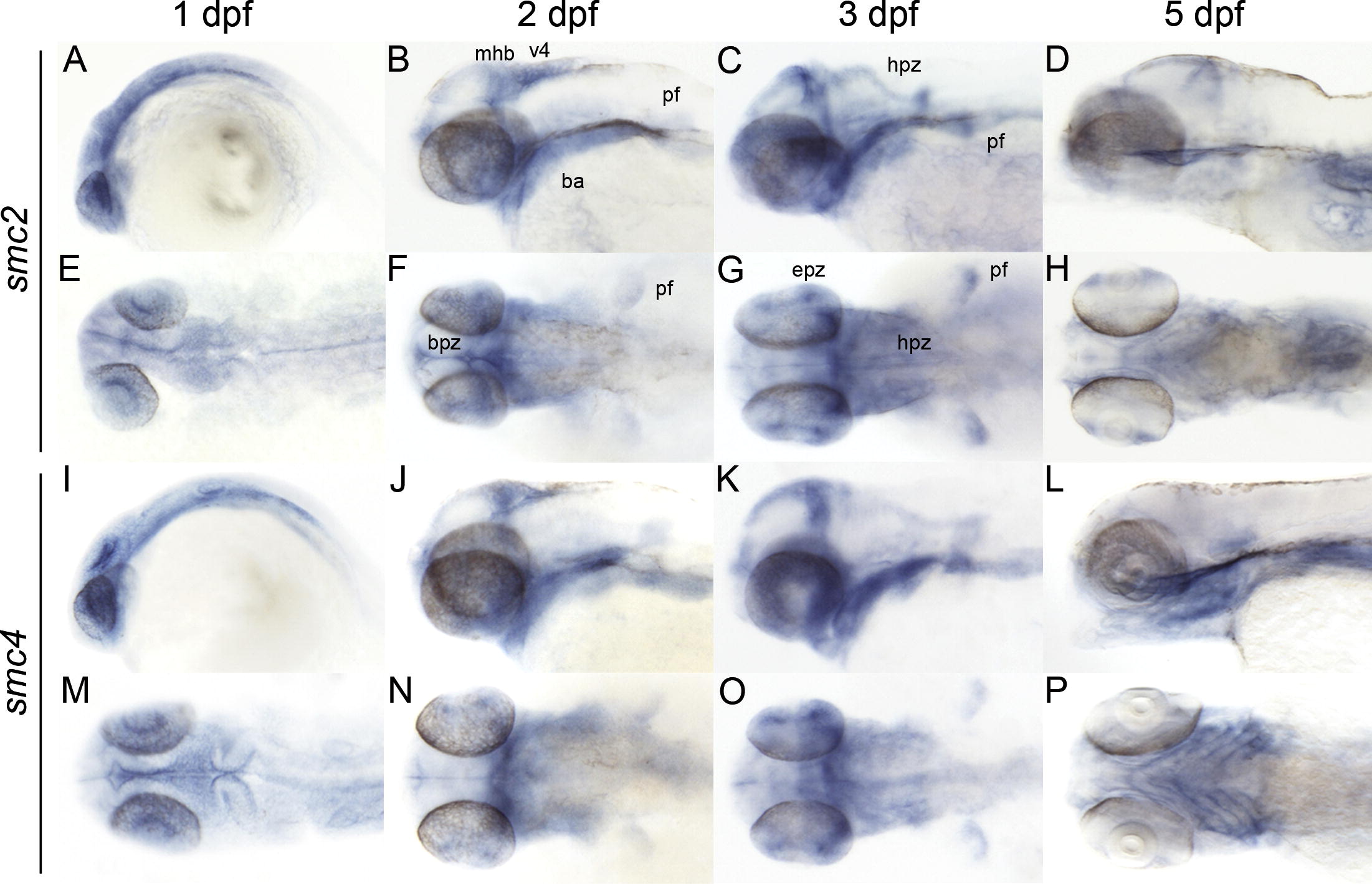
Fig. 3 Late embryonic and larval expression (1–5 dpf) of zebrafish condensin subunits. Whole mount in situ hybridization of zebrafish at the indicated stage with antisense riboprobes smc2 (A–H), and smc4 (I–P). A–D, I–L are lateral views. E–H, M–P, are dorsal views. Anterior is to the left for all. ba, branchial arches; bvz, brain ventricular zone; epz, proliferative zone in the eye; hpz, hindbrain proliferative zone; mhb, midbrain hindbrain boundary; ov, otic vesicle; pf, pectoral fin; v4, 4th ventricle.
Figure Data
Acknowledgments
This image is the copyrighted work of the attributed author or publisher, and
ZFIN has permission only to display this image to its users.
Additional permissions should be obtained from the applicable author or publisher of the image.
Reprinted from Gene expression patterns : GEP, 9(8), Mönnich, M., Banks, S., Eccles, M., Dickinson, E., and Horsfield, J., Expression of cohesin and condensin genes during zebrafish development supports a non-proliferative role for cohesin, 586-594, Copyright (2009) with permission from Elsevier. Full text @ Gene Expr. Patterns